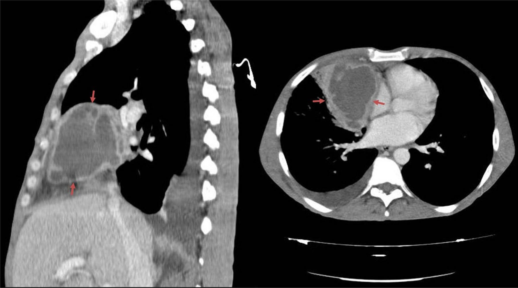
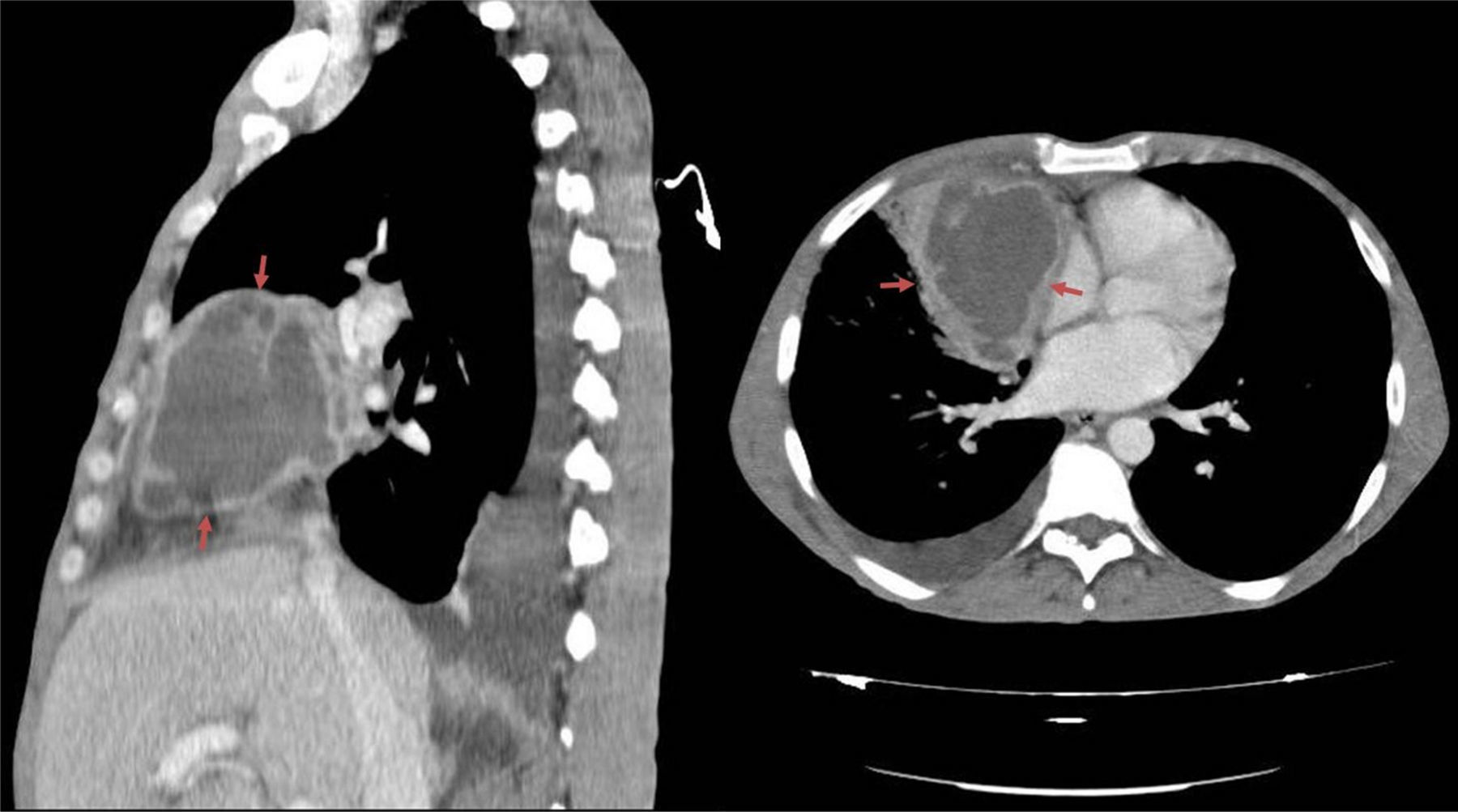
A 51-year-old man, former smoker with a history of hepatitis C infection, presented with symptoms of fever, arthralgia, and weight loss. After case history and physical examination were completed, which were unremarkable, additional tests were requested: complete blood count, serum biochemistry, viral serologies, autoimmunity, Mantoux and blood cultures. Of note was C-reactive protein 238mg/l. Imaging studies were performed, and a chest radiograph revealed a picture of water density in the right hilum and the chest computed tomography (CT) showed an anterior mediastinal mass 9×7cm consistent with infected embryonic remnants (Fig. 1). The patient was diagnosed with an infected pleuropericardial cyst, and antibiotic treatment began. Progress was good so the patient was discharged and scheduled for surgical extraction of the cyst.
Pleuropericardial cyst is an uncommon lesion that occurs at an incidence of 1 in 100,000 cases. It is normally located in the mid-mediastinum, and is often asymptomatic and found by chance,1 but it may be complicated by compression from adjacent structures,2 infection or bleeding from the cyst itself.1 Treatment is conservative, and surgical treatment is reserved for symptomatic cases. In our patient, both the form of presentation (infection) and the site (anterior) are unusual.
Please cite this article as: Yeste Martín R, Navarro Marín LJ, Herrero Rodríguez, C. Quiste pleuropericárdico: localización y presentación inusuales. Arch Bronconeumol. 2017;53:208.