
A 70-year-old ex-smoker man presented with low-grade fever and progressive dyspnea. His past medical history was significant for chronic obstructive pulmonary disease, combined pulmonary fibrosis and emphysema syndrome, and chronic pulmonary aspergillosis (CPA) secondary to previous tuberculosis (Fig. 1). A chest radiograph showed a stable destructive pattern of the right lung but disappearance of a previously documented aspergilloma within a right upper lobe cavity (Fig. 2A). A contrast-enhanced thoracic computed tomography (CT) confirmed the vanishing of the aspergilloma but also demonstrated the appearance of a new pleural effusion and an enhancing thickened pleural surface of the right hemithorax (Fig. 2B). A thoracentesis confirmed a fungal empyema, and a chest “pig-tail” catheter was inserted (Fig. 2C). The patient was started on voriconazole (200mg twice daily) and has shown, six months later, an excellent response with resolution of his symptoms and of the pleural effusion.
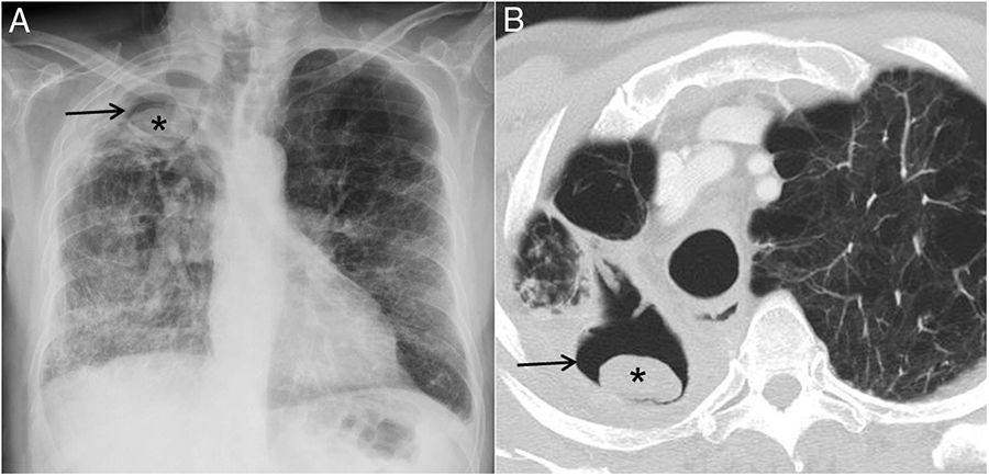
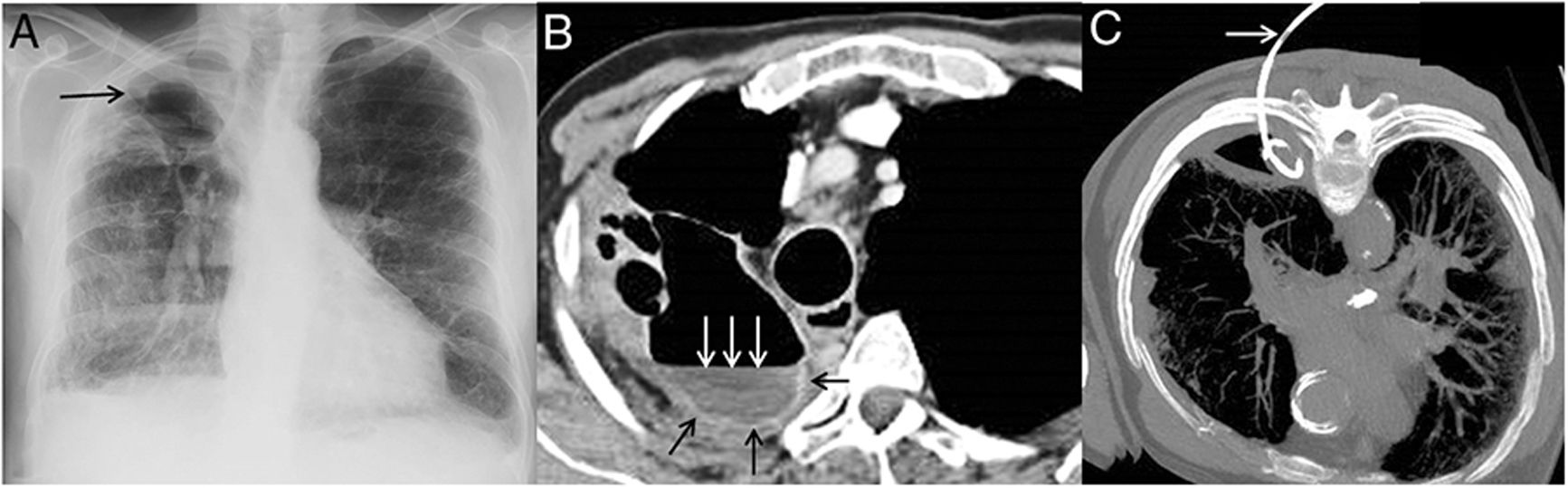
Chest radiograph (A) shows disappearance of the previously documented aspergilloma (arrow). Axial contrast-enhanced CT (B) confirms the vanishing aspergilloma; note the presence of an air-fluid level (white arrows) within the right upper hemithorax and the enhancing pleural surface (black arrows) suggesting an empyema. Axial CT image (C, the patient is in the prone decubitus position) shows a pig-tail catheter within the right pleural space (arrow).
CPA is an uncommon but problematic pulmonary disease most commonly complicating previous tuberculosis infections. The most common forms of CPA are chronic cavitary pulmonary aspergillosis and aspergilloma. Chest radiographs and CT remain the most important imaging modalities for the suspicion and diagnosis of CPA. Spontaneous vanishing of a pulmonary aspergilloma is an extremely rare phenomenon with only three previous described case reports in the literature. Disappearance of a previously documented aspergilloma on imaging should prompt physicians to suspect a spontaneous perforation of the pleura and appearance of a fungal empyema, especially in patients with CPA.
Author's contributionThe authors have made substantial contributions regarding not only the conception and design of the manuscript, but also the drafting and critical revision of the article.
Conflict of interest and FundingNone of declare.


