


Pulmonary nocardiosis (PN) is a severe infection with a high morbidity and mortality that mainly affects immunocompromised patients. In recent years, an increase in PN cases has been detected among patients with chronic obstructive pulmonary disease (COPD). The factors that are associated with its presence and determine its prognosis remain unknown.
MethodsRetrospective study of COPD patients diagnosed with PN over the period from 1997 to 2009 at the Hospital de la Santa Creu i Sant Pau, in Barcelona (Spain). Demographic, clinical, microbiological and evolution data were evaluated in all cases.
ResultsThirty patients were identified with PN and COPD. Mean age (standard deviation) was 76 (7) years and the mean FEV1 was 40 (14)%. Chronic respiratory failure was observed in 56.7% patients and 51.7% had received systemic corticosteroid therapy previous to the PN diagnosis. The most common symptoms were cough and dyspnea (90%). Alveolar infiltrates were observed in 60% of the cases. The most frequently isolated Nocardia species was N. cyriacigeorgica (68%). The one-month mortality rate was 17%, while the one-year mortality rate was 33%. The factors associated with mortality within the first year included previous systemic corticosteroid treatment, less than three months of specific antibiotic therapy and active associated neoplasm.
ConclusionsPN affects patients with moderate-severe COPD and has high short- and mid-term mortality rates. Previous corticosteroid treatment, specific antibiotic therapy for less than 3 months and active neoplasia were factors associated with mortality.
La nocardiosis pulmonar (NP) es una infección grave que afecta principalmente a pacientes inmunodeprimidos y se asocia a una elevada morbimortalidad. En los últimos años se ha detectado un aumento de casos de NP en pacientes con enfermedad pulmonar obstructiva crónica (EPOC). Los factores que se asocian a su presencia y determinan su pronóstico son desconocidos.
Material y métodosEstudio retrospectivo de los pacientes con NP y EPOC diagnosticados durante el período 1997–2009 en el Hospital de Sant Pau de Barcelona. Se recogieron datos demográficos, clínicos, microbiológicos y evolutivos.
ResultadosSe identificaron 30 pacientes con NP y EPOC. La media (desviación estándar) de edad fue de 76 (7) años, y la de FEV1, del 40 (14)%. El 56,7% presentaba insuficiencia respiratoria crónica y el 51,7% había recibido tratamiento corticoesteroideo sistémico previo al diagnóstico. Los síntomas más frecuentes fueron tos y disnea (90%). En el 60% de los casos se observaron infiltrados alveolares. La especie de Nocardia aislada con mayor frecuencia fue N. cyriacigeorgica (68%). La mortalidad el primer mes fue del 17%, y al año, del 33%. Los factores que se asociaron con mortalidad al año fueron tratamiento corticoesteroideo sistémico previo, antibioticoterapia específica durante menos de 3 meses y presencia de neoplasia activa asociada.
ConclusionesLa NP puede afectar a pacientes con EPOC moderada y grave y cursa con una elevada mortalidad a corto y medio plazo. El tratamiento corticoesteroideo previo, recibir una terapia antibiótica específica durante menos de 3 meses y la presencia de neoplasia activa son los factores que se asocian a la mortalidad.
Pulmonary nocardiosis (PN) is the most frequent from of presentation of infection due to Nocardia spp. in humans.1 Its main risk factor is cellular immunosuppression, which mainly affects patients with hematologic diseases, solid organ transplant, those infected with human immunodeficiency virus (HIV) and patients who receive prolonged treatments with corticosteroids, cytotoxic drugs and/or biological therapies with anti-TNF.1–5 Its associated mortality is very high, ranging from 14% to 40%,1 increasing to 60%–100% in cases in which there is dissemination to the central nervous system (CNS).1,6,7
In recent years, there has been evidence of an increase in the number of cases of PN in patients with Chronic Obstructive Pulmonary Disease (COPD). An initial study by Menéndez et al. that evaluated 10 patients affected by PN showed that COPD was an important risk factor for developing the disease, even greater than having a neoplastic disease or being an HIV carrier.6 Later studies confirmed this observation. In 31 cases of PN,2 Martínez et al. observed that COPD was the third most frequent risk factor, surpassed only by chronic steroid treatment and solid organ transplantation. Likewise, in addition to confirming that lung affectation was the most frequent form of presentation of Nocardiosis in humans, other recent studies3,6 observed that the most prevalent underlying conditions were HIV infection3 and the presence of chronic respiratory diseases like COPD.6,8
All these findings suggest that COPD, especially in advanced stages, can play a very important role in the pathogeny of PN. COPD is a disease that has a great impact in our setting, affecting almost 10% of the population between the ages of 40 and 70, and it is already the third cause of hospitalization and the fourth overall cause of mortality. This mortality rate increases year after year, and it is predicted to become the third cause of death by the year 2020.9,10 Due to this reason, it is expected that PN in COPD patients will be seen more and more frequently and will acquire great importance due to its potential impact on the prognosis of the disease.
To date, no studies have evaluated either the factors that associate PN in COPD patients or the prognosis of the disease in this group of patients. The objective of the present study is to describe the characteristics of COPD patients who present with PN and to know those factors that are associated with its prognosis.
MethodsA retrospective study including all the COPD patients diagnosed with PN at Hospital de la Santa Creu i Sant Pau in Barcelona from 1997 to 2009.
DefinitionsThe diagnosis of pulmonary nocardiosis was established by the presence of symptoms of respiratory infection and the isolation of Nocardia spp. in the cultures of respiratory samples.
An infection was considered to be disseminated when Nocardia spp. was isolated in a hemoculture or when there was a demonstrated infection in other organs and/or in subcutaneous cell tissue simultaneous to the lung infection.
The diagnosis of COPD was established according to current national clinical practice guidelines.11
Microbiological IdentificationFor the diagnosis of pulmonary nocardiosis, samples were taken from sputum, tracheal aspiration or bronchoalveolar lavage.
The samples were examined microscopically after Gram staining in order to determine the presence of microorganisms and to establish the quality of the sputum and tracheal aspirate samples.12
The cultures were done conventionally in blood agar, chocolate agar, EMB agar and BCYE agar media that were incubated at 37°C in an aerobic atmosphere with 5% of carbon dioxide for 2–7 days.
The presumptive identification of Nocardia spp. was done based on the microscopic characteristics of the isolations and the macroscopic morphology of the colonies: branching Gram-positive bacilli, partially acid-fast with modified Kinyoun stain, that presented hyphae on the surface of the colony.13,14
The identification at the species level was done by molecular analysis, by polymerase chain reaction (PCR) and partial sequencing of a fragment of 800pb of the 16S rDNA gene and later comparison of the sequences with those deposited in the GenBank. Nocardia abscessus and Nocardia asiatica were differentiated by PCR and secA1 gene sequencing.15
Study VariablesData were collected for demographics (age, sex, body mass index [BMI]), comorbidities (arterial hypertension, diabetes mellitus, dyslipidemia, renal failure, hepatopathy and neoplasm), other respiratory diseases, lung function, presence of chronic respiratory failure and data about immunological state (presence of hematologic disease, human immunodeficiency virus infection, solid organ transplant, immunosuppressant treatment). Other data collected were clinical (presentation: cough, dyspnea, fever, leukocytosis, respiratory failure, dissemination to other organs/CNS affectation), radiological using chest radiography and/or chest CT (alveolar infiltrates, cavitation, pleural effusion); microbiological (isolated Nocardia spp. species) and the duration of the treatment received for Nocardiosis. Finally, mortality was assessed both 30 days and 1 year after diagnosis.
Statistical AnalysisThe statistical analysis was carried out with SPSS software (V 17.0).
According to the nature of the variables involved, we evaluated their association with the main variable, “Mortality” (after 30 days and 1 year). For the categorical variables, a bivariable study was completed with contingency tables and the inference with the Chi-squared or Fisher's exact tests. The quantitative variables are reported with the mean value and standard deviation, and the inference was done with the Student's t test for independent data. The level of significance used was 5% (bilateral approximation).
ResultsCharacteristics of the IndividualsIncluded in the analysis were the 30 patients with COPD and PN who were identified during the period studied. Mean age (standard deviation) was 76 (7), mean BMI was 25 (3)kg/m2 and the distribution by sex was 28 men (93.3%) and 2 women (6.7%). Twenty-eight were ex-smokers (93.3%) and 2 (6.7%) continued to smoke actively at the time of infection. The most frequent comorbidity was arterial hypertension, present in 12 of the subjects (40%), followed by dyslipidemia in 7 patients (23.3%) and presence of active neoplasm, which was observed in 7 cases (23.3%), 5 of these being pulmonary in origin and 2 extrapulmonary. Four patients (13.3%) presented kidney failure, 3 (10%) had diabetes mellitus and 1 chronic hepatopathy (3.3%). Four or more comorbidities were detected in only 2 (6.7%) patients at the time of diagnosis. Mean FEV1 was 40% (14%) and 17 cases (56.7%) presented chronic respiratory failure at the time of diagnosis. Fifteen subjects (51.7%) had received corticosteroid treatment prior to the infection and 5 (16.7%) took corticosteroids regularly. Immunosuppression was found in 7 patients (23.3%), 3 of whom had undergone solid organ transplantation and 1 had received chemotherapy for active malignant neoplasm. The rest received chronic corticosteroid treatment due to other indications. None of the patients presented positive serology for HIV (evaluated in 10 patients) nor had they received biologic treatment with anti-TNF.
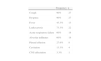
Symptoms, Lab and Radiology ResultsThe most frequently observed symptoms were cough and dyspnea in 27 patients (90%) and associated fever in 13 (43.3%). In one case, dissemination to the central nervous system was detected. Blood work-up confirmed leukocytosis in 22 cases (73.3%); arterial blood gas analyses showed that 18 (60%) presented acute or chronic exacerbation of respiratory failure. Radiography showed evidence of parenchymatous affectation in the form of alveolar infiltrates in 18 (60%), cavitation in 4 (13.3%) and pleural effusion in 8 (27.6%) cases (Table 1).
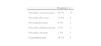
Microbiological CharacteristicsThe isolated species of Nocardia spp. are shown in Table 2. The species Nocardia cyriacigeorgica was the most frequent and was isolated in 17 patients (56.7%).
The patients received specific antibiotic treatment for nocardiosis during an average of 4 (3) months.
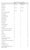
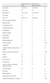
Prognostic FactorsWithin the first month, the mortality rate was 17% and 33% within the first year of diagnosis. No statistically significant differences were detected among the characteristics evaluated in the group of patients who died within the first month compared with those who still survived (Table 3). In contrast, in the same comparison done after 12 months, it was observed that the group of patients who did not survive had a significantly greater incidence of active neoplasms, had received more systemic corticosteroids and had not received prolonged antibiotic treatment (more than three months) to treat their PN (Table 4).
Prognostic Factors One Month after Diagnosis of Pulmonary Nocardiosis.
| Group of Survivors (n=25) | Group of Non-Survivors (n=5) | P | |
| Age, yearsa | 75.7 (7.2) | 75.8 (10.8) | ns |
| Sex, males | 23 | 5 | ns |
| BMI, kg/m2a | 24.6 (4.1) | 25.5 (0.83) | ns |
| Ex-smoker | 24 | 4 | ns |
| FEV1, %a | 38.9 (15) | 46.6 (5.6) | ns |
| Chronic respiratory failure | 14 | 3 | ns |
| Bronchiectasis | 5 | 1 | ns |
| Comorbidities (≥4) | 1 | 1 | ns |
| Arterial hypertension | 10 | 2 | ns |
| Dyslipidemia | 6 | 1 | ns |
| Diabetes mellitus | 3 | 0 | ns |
| Renal failure | 3 | 1 | ns |
| Hepatopathy | 1 | 0 | ns |
| Immunosuppression | 6 | 1 | ns |
| Active neoplasm | 4 | 3 | ns |
| Alcoholism | 0 | 1 | ns |
| Antibiotic therapy the month before | 12 | 3 | ns |
| Recent systemic corticosteroids | 12 | 3 | ns |
| Cough | 23 | 4 | ns |
| Dyspnea | 22 | 5 | ns |
| Fever (≥38°C) | 11 | 2 | ns |
| Leukocytosis | 19 | 3 | ns |
| Acute or chronic exacerbated respiratory failure | 14 | 4 | ns |
| Alveolar pattern in thoracic radiology | 15 | 3 | ns |
| Cavitation | 3 | 1 | ns |
| Pleural effusion | 7 | 1 | ns |
| Dissemination to CNS | 0 | 1 | ns |
| Specific antibiotic treatment for PN during >3 months | 9 | 0 | ns |
BMI: body mass index; FEV1: forced expiratory volume in 1s; CNS: central nervous system; ns: not statistically significant.
Prognostic Factors 12 Months After Diagnosis of Pulmonary Nocardiosis.
| Group of Survivors (n=20) | Group of Non-Survivors (n=10) | P | |
| Age, yearsa | 75.20 (6.5) | 76.20 (9.9) | ns |
| Sex, males | 18 | 10 | ns |
| BMI, kg/m2a | 24.15 (3.85) | 25.94 (3.99) | ns |
| Ex-smoker | 19 | 9 | ns |
| FEV1, %a | 36.95 (15.1) | 46.60 (9.9) | ns |
| Chronic respiratory failure | 13 | 4 | ns |
| Bronchiectasis | 5 | 1 | ns |
| Comorbidities (≥4) | 1 | 1 | ns |
| Arterial hypertension | 9 | 3 | ns |
| Dyslipidemia | 6 | 1 | ns |
| Diabetes mellitus | 3 | 0 | ns |
| Renal failure | 3 | 1 | ns |
| Hepatopathy | 0 | 1 | ns |
| Immunosuppression | 4 | 3 | ns |
| Active neoplasm | 1 | 6 | .002 |
| Alcoholism | 0 | 1 | ns |
| Antibiotic therapy in the previous month | 9 | 6 | ns |
| Recent systemic corticosteroids | 7 | 8 | .049 |
| Cough | 19 | 8 | ns |
| Dyspnea | 19 | 8 | ns |
| Fever (≥38°C) | 9 | 4 | ns |
| Leukocytosis | 16 | 6 | ns |
| Acute or chronic exacerbated respiratory failure | 13 | 5 | ns |
| Alveolar pattern | 12 | 6 | ns |
| Cavitation | 2 | 2 | ns |
| Pleural effusion | 4 | 4 | ns |
| Dissemination to the CNS | 0 | 1 | ns |
| Antibiotic treatment for >3 months | 9 | 0 | .048 |
BMI: body mass index; FEV1: forced expiratory volume in 1s; CNS: central nervous system; ns: not statistically significant.
The present study describes the characteristics of the patients with COPD who presented with PN and the factors that are associated with a poor prognosis of the disease. Until now, such data have not been published, and given the high prevalence of COPD and the potential impact of PN, they may greatly influence daily clinical practice.
The most important findings are that PN in COPD patients mainly affects those with advanced lung disease who receive systemic corticosteroid treatment, that its associated mortality is very high and that the presence of active neoplasm, antibiotic treatment for less than 3 months and systemic corticosteroid therapy are factors that are related with mortality 12 months after the diagnosis of PN.
The characteristics of the COPD patients studied demonstrate some important differences when compared with data from non-COPD patients with PN that had been previously reported. The most notable characteristic of our group is that it affects patients with moderate-intense lung function alteration, with the presence of chronic respiratory failure in more than half of the patients. To date, no other study had analyzed lung function in patients with PN. Another important finding is that more than half of the patients had received systemic corticosteroid treatment prior to diagnosis. This datum coincides with previous studies that related steroid treatment with the presence of PN in different patient populations.1,2,6,14,16
From the rest of the variables studied, we have observed a marked difference in the incidence between sexes, with a high predominance among men, which is probably explainable by the lower prevalence among women of smoking history and COPD diagnosis.9,10 It is also striking that less than 5% of the patients present a high number of associated comorbidities (4 or more), reinforcing the finding that PN is more associated with advanced pulmonary pathology and not a more general multi-pathological state.
From a clinical standpoint, the presentation of PN in COPD patients is very unspecific. Cough and dyspnea are the most frequent symptoms, and there is a lack of fever in more than half of the cases, unlike in other PN patient populations where studies have shown that fever was a frequent symptom.1,2,17 Likewise, the presence of COPD may favor a predominance of respiratory symptoms over systemic ones, a fact that was confirmed in a former study of 10 PN cases where cough was also the most frequent symptom in patients with chronic pneumopathies.6
In this group of patients, no specific radiological data for the presence of PN have been found. The most common form of presentation was alveolar infiltrates, coinciding with the study by Martínez et al.2 Cavitation, the most characteristic radiological finding reported in previous studies in immunosuppressed patients and PN,18,19 was only found in approximately 10% of the COPD patients.
As for microbiology, the species of Nocardia that was most frequently isolated associated with COPD was N. cyriacigeorgica (56.7%). This contrasts with previous studies in other patient groups where the most frequently isolated species were Nocardia asteroides,2,20,21Nocardia farnicia8 and Nocardia nova complex,22 although these results are difficult to compare due to the recent taxonomic changes in the genus Nocardia.13,23
The overall mortality one year after diagnosis with PN was 33%, which is considerably high when compared with other previous studies of COPD patients who presented exacerbations requiring hospitalization, where mortalities of 22%24 and 23%25 were detected 12 months after the exacerbation.
From a prognostic standpoint, three factors have been related with mortality one year after diagnosis. First of all, the presence of active neoplasm, a fact that was expected and can justify both an altered baseline immunological state as well as being a direct cause of demise, a fact that has not been analyzed in this present study. The second factor is the presence of treatment with systemic corticosteroids prior to the diagnosis of the infection. It is well known that corticosteroids, through their anti-inflammatory genomic mechanism of action,26 modulate the inflammatory response of the host against germs, and that its chronic administration may be the cause of immunosuppression and associated severe infections,27 a circumstance that could explain this finding.
Finally, it has been reported that antibiotic treatment directed at Nocardia administered for less than 3 months is associated with greater one-year mortality after diagnosis. The current clinical practice guidelines about the treatment of the disease28 are not clear in these cases. They recommend treatment for 12 months when there is affectation of the CNS and immunosuppression and for 3 months in the cases of cutaneous nocardiosis, but there is no clear indication for these patients. This finding can reinforce the need for prolonged treatment in cases of lung affectation with Nocardia spp., regardless of the immunological state of the patient and whether the affectation is only bronchial or parenchymatous.
It is important to underline that, in most prior studies about PN,1,6–8 the factor that was most related with mortality was dissemination of Nocardia to the CNS, a fact that occurred in only one patient in our study group. This demonstrates once again that the behavior of Nocardia spp. in patients with COPD differs from its behavior in other risk groups.
It should be emphasized that this present study has some limitations that should be considered. In the first place, it is a retrospective study in which many potential biases have not been controlled. The number of patients studied is too small to be able to perform a multivariate analysis, correct for multiple variables or reach more solid conclusions. This is despite the fact that it is a very similar number to all the previously published studies about PN,3,6 and that it is the most numerous study with patients with PN and lung pathology published to date. One of the main factors that have limited the inclusion of patients has been the required spirometry for the diagnosis of COPD, as indicated in current clinical practice guidelines.11 Another limitation is that mortality was studied globally, not only mortality attributable to PN; thus, in some cases mortality may have been due to pathologies unrelated with PN. Finally, other important limitations of the study are that we do not know the exact dosage of corticosteroids administered or the stage of the active neoplasms, which are factors related with mortality. In addition, we lacked chest CT for all the patients to better understand the presence or absence of alterations in the pulmonary parenchyma.
The main conclusions of this study are that PN is a severe disease with a high short- and mid-term mortality rate in COPD patients. Its presentation is varied and especially affects patients with moderate-intense alteration of the lung function, chronic respiratory failure and recent systemic corticosteroid treatment. The presence of active neoplasm, recent systemic corticosteroid treatment and antibiotic treatment during a period of less than 3 months are the factors associated with poor prognosis and with mortality 12 months after the diagnosis of PN. Although extensive prospective studies are necessary to validate these findings, these results may promote the active, early identification of the bacteria in this type of patients, while trying to avoid adjuvant corticosteroid treatment as well as maintaining antibiotic treatment aimed at Nocardia for more than 3 months in order to improve prognosis.
Conflict of InterestThe authors declare no conflict of interest.
Please cite this article as: Garcia-Bellmunt L, et al. Nocardiosis pulmonar en pacientes con EPOC: características y factores pronósticos. Arch Bronconeumol. 2012;48:280–5.


