

To the editor: The genus Nocardia includes various species that have been implicated in human infection. N asteroides is the species most frequently isolated in patients with infection by Nocardia. During a recent taxonomical review, 2 new species were added to this genus: N farcinica and N nova.1 Below we describe a case of pulmonary disease due to N nova which contributes to the small body of literature on the role of this microorganism in pulmonary infections.2-4
The patient was a 78-year-old man with a personal history of rheumatic polymyalgia undergoing treatment with 10 mg/day of oral corticosteroids for more than 9 months. He also met clinical criteria for chronic bronchitis, with grade II/IV dyspnea in stable condition. He came to our hospital complaining of dyspnea with minimal effort which had steadily increased over the 4 weeks prior to admission. He also showed signs of orthopnea and paroxysmal nighttime dyspnea. Up to 90 mg/day of oral prednisone was prescribed to treat the increase in dyspnea and the rheumatic polymyalgia. The patient reported having cough and purulent sputum as well as a fever of around 38 ºC on the days prior to admission. On physical examination the patient was alert and oriented; his blood pressure was 160/90 mm Hg and temperature was 38 ºC. Dependent edema with pitting was noted along the full length of the lower extremities (including the groin). Basal crackles on both sides, best heard over the lower third of the right hemithorax, were detected on pulmonary auscultation. Respiratory rate was 24 breaths/min. Arterial blood gas analysis showed an arterial oxygen pressure of 55 mm Hg, an arterial carbon dioxide pressure of 30 mm Hg, a pH of 7.44, and oxygen saturation at 89%. A chest x-ray showed alveolar condensation in the right lower lobe, and multiple nodular images in both lower lobes, some containing ill-defined radiolucent areas. The patient was initially treated with a third-generation cephalosporin and amikacin. Response was poor in the first 24 to 48 hours, and the patient developed severe heart failure, with some improvement following diuretic treatment. Respiratory insufficiency persisted and an inspiratory oxygen fraction of approximately 0.35% to 0.40% was required. Sputum Gram staining revealed gram-positive branching bacilli. Acid-fast bacteria smear tests and Löwenstein-Jensen cultures were negative for mycobacteria; however, Nocardia, identified biochemically (resistance to lysozymes, sensitivity to ampicillin and erythromycin among others) as N nova, grew in both aerobic and mycobacterial cultures. Identification was completed through analysis of gene hsp65 with primers Tb11 (5'-ACCAACGATGGTGTGTCCAT) and Tb12 (5'-CTTGTCGAACCGCATACCCT). Forty-five cycles of amplification were performed (1 minute at 94 ºC, 1 minute at 60 ºC, and 1 minute at 72 ºC), with a final extension of 10 minutes at 72 ºC.5 Later, a restriction analysis was carried out with Msp1 and Hinf1 enzymes. No restriction site was obtained with Hinf1, which gained a single band of 439 bp. Msp1 enzyme obtained 3 bands of 130, 110 and 75 bp. According to published data, the profile obtained coincided with that of N nova (Figure). Once Nocardia was identified, specific antibiotic treatment with trimethoprim-sulfamethoxazole was initiated, and the patient responded favorably. At discharge, edema persisted although it had gradually decreased in the last days of his stay. He was afebrile and chest x-rays revealed satisfactory improvement.
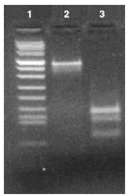
Figure. Profile of bands obtained by polymerase chain reaction/restriction fragment length polymorphism of gene hsp65 using Msp1 and Hinf1 restriction enzymes. Lane 1: molecular weight marker VIII (Roche); lane 2: restriction fragment using Hinf1 enzyme; lane 3: restriction fragment using Msp1 enzyme.
N nova was first isolated and described by Tsukamura6 in 1982. This group is mainly characterized by its sensitivity to ampicillin and erythromycin.6,7N nova has been described as the cause of skin infection and sinusitis, as well as infection through catheters. Three cases of pneumonia caused by N nova have also been reported: one after a heart transplant coinfected with Aspergillus fumigatus,2 another in a patient with lung cancer,3 and a third case, similar to the one presented here, of a 52-year-old Japanese woman previously treated with steroids and cyclophosphamide for polymyositis.4 The application of appropriate identification methods, among them techniques from molecular biology, increases the number of isolates of this identified microorganism.


