


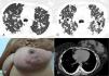
A 31-year-old woman was admitted with complaints of weight loss, dry cough, and fever. Chest computed tomography (CT) revealed small confluent nodules in both lungs (Fig. 1a). The patient returned for consultation six months later, with worsening of her general condition. She reported the appearance of painful palpable nodules in her left breast 2 months previously, with productive cough and 15kg weight loss in the previous 6 months. Physical examination confirmed nodules in the left breast (Fig. 1b). Laboratory tests were normal and an HIV test was negative. Magnetic resonance imaging of the breasts suggested the presence of abscesses. A new CT showed multiple cavitating masses in the upper regions of both lungs (Fig. 1c) and lesions in the left breast (Fig. 1d). Three sputum samples were positive for acid-fast bacilli. Cultures of breast aspirate and sputum were positive for Mycobacterium tuberculosis. The final diagnosis was pulmonary and breast TB.

High-resolution computed tomography of the upper lobes (A) revealed small confluent nodules in both lungs. Physical examination of the left breast (B) showed palpable and visible lumps in the inner quadrants. The right breast was normal. Follow-up computed tomography at the same level (C) demonstrated worsening of the pulmonary lesions, with the appearance of multiple cavitating masses in the upper regions of both lungs. A chest computed tomography with mediastinal window setting showed the lesions in the left breast (D).
TB is a major health problem in underdeveloped and developing countries, and its incidence in the developed world is increasing. Nevertheless, breast TB remains rare. The gold standard for the diagnosis of TB is detection of M. tuberculosis by Ziehl–Neelsen staining or culture.1,2
Please cite this article as: Bazi-Fontes F, Zanetti G, Marchiori E. Tuberculosis pulmonar y mamaria: una asociación inusual. Arch Bronconeumol. 2015;51:598–599.


