
The objective of this present study was to describe the clinical-radiological and lung function characteristics of hypersensitivity pneumonitis due to exposure to isocyanates (HPI). Included for study were all those patients diagnosed with HPI (n=5) from 1995 to 2010. In all cases, chest CT and complete lung function studies were done. The diagnosis was made with positive specific bronchial provocation tests (BPT). A predominance of males and pattern variability in the radiological alterations were observed. The BPT was positive due to a decline in FVC or DLCO in all cases except in one patient in whom the diagnosis was established with clinical criteria. Positive IgG to isocyanates was confirmed in only one patient. The radiological presentation of HPI may be different from the more common HP in our setting and run their course with less clinical affectation on the BPT.
El objetivo del presente trabajo fue describir las características clínico-radiológicas y de función pulmonar de la neumonitis por hipersensibilidad por exposición a isocianatos (NHI). Se estudiaron todos los pacientes con diagnóstico de NHI (n=5) durante los años 1995-2010. En todos ellos se realizó TC torácico y estudio de función pulmonar completo. El diagnóstico se realizó mediante prueba de provocación bronquial específica (PPBE) positiva. Se observó un predominio de varones y una variabilidad de patrones en las alteraciones radiológicas. La PPBE fue positiva por descenso de la FVC o de la DLCO en todos los casos excepto en un paciente en que se estableció por criterios clínicos. Tan solo en un paciente se constató una IgG positiva a isocianatos. La presentación radiológica de la NHI puede ser distinta de las NH más frecuentes en nuestro medio y cursan con menor afectación clínica en la PPBE.
Hypersensitivity pneumonitis (HP) is an interstitial disease of known etiology.1 Currently, more than 50 antigens have been identified as being able to induce HP, and the majority of them are organic, high-molecular-weight compounds. Although it is uncommon, inorganic low-molecular-weight compounds have also been implicated in the genesis of HP, the most characteristic example of this being isocyanates.2
Isocyanates are chemical compounds that are able to polymerize flexible or rigid substances, making them widely used in the fabrication of varnishes, plastics, insulating material, motor vehicle upholstery, lacquers, insecticides, etc. They are the most frequent causal agent identified in occupational asthma,3 and they have also been identified, albeit rarely, as a cause of lung parenchyma affectation. Although its pathogenesis is unknown, the presence of systemic symptoms, lymphocytosis in the bronchoalveolar lavage and high levels of specific IgG have suggested that the cause of this pathology may be a mechanism similar to HP caused by organic compounds.4,5
In this study, we described 5 cases of hypersensitivity pneumonitis due to exposure to isocyanates (HPI) in which the patients developed the disease after exposure to isocyanates at their place of work. This is the largest such series described in our country.
Case ReportA retrospective study of cases and controls analyzed all those patients diagnosed with HPI from 1995 to 2010 who were referred to an occupational respiratory pathology unit at a tertiary hospital for diagnostic confirmation using specific bronchial provocation tests (SBPT) (n=5). Based on their medical files, we analyzed clinical characteristics, including physical exploration, analytical determinations, thoracic computed tomography (CT) and lung function studies, including spirometry, static lung volumes, CO diffusing capacity test, methacholine challenge and bronchoscopy results in one of the patients.
Lung function tests were done in accordance with European Respiratory Society (ERS) guidelines with a MasterLab device (MasterLab, Jaegger, Germany).6,7 The spirometry reference values used were for Mediterranean populations.8 Lung volumes were measured with plethysmography, while the single-breath method was used for carbon monoxide diffusing capacity. In both cases, the predicted values used were those proposed by the ERS.6,7 The non-specific bronchial provocation test with methacholine was carried out with the method described by Chai et al.9 The test was considered negative if the PC20 FEV1 was greater than 16mg/ml, in accordance with ATS criteria.10
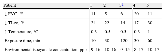
The patients were exposed to toluene diisocyanate (TDI) in a controlled provocation booth, as previously described,11 controlling that the level of isocyanates did not surpass 20ppb at any time by means of a MDA 7100 device (MDA® Scientific, Inc., Glenview, IL, USA). The patients were initially exposed to a placebo for 15min and later, on successive days, to increasingly longer times up to a maximum of 2h. For all patients, we measured temperature, FEV1, FVC and DLCO after 20min of exposure and every hour for the following 8h. The test was considered positive if there was a decrease of >15% in FVC and/or a decrease >20% in DLCO or a decrease in FVC between 10% and 15% together with an increase in temperature >0.5°C. The test could also be considered positive if a minimum of 3 of the following clinical criteria were observed without detecting changes in lung function studies: temperature >37°C, leukocytosis, radiological changes, lung auscultation changes, dyspnea and/or arthromyalgia.12 All patients gave their written informed consent for the SBPT.
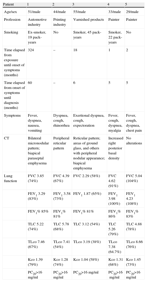
The clinical, radiological and lung function characteristics of the patients are shown in Table 1. The immunoglobulin determinations carried out with ELISA were negative except in patient number 2, who presented positive IgG for isocyanates. SBPT was positive in all cases with the criteria of decreased FVC or DLCO, except in one patient in whom positivity was established by clinical criteria (Table 2). Bronchoscopy was carried out in 4 patients, although it was only possible to obtain data for patient number 2. The presence of lymphocytes in the bronchoalveolar lavage of said patient was 39%, and a pathology sample obtained by transbronchial biopsy showed mature collagen fibrosis, areas of honeycomb pattern with macrophages within the air spaces and a moderate centrilobular inflammatory component, plasma cells, histiocytes and some eosinophils.
Clinical, Radiological and Lung Function Characteristics of the Patients.
| Patient | 1 | 2 | 3 | 4 | 5 |
| Age/sex | 51/male | 44/male | 55/male | 33/male | 29/male |
| Profession | Automotive industry | Printing industry | Varnished products | Painter | Painter |
| Smoking | Ex-smoker, 19 pack-years | No | Smoker, 45 pack-years | Smoker, 22 pack-years | No |
| Time elapsed from exposure until onset of symptoms (months) | 324 | – | 18 | 1 | 2 |
| Time elapsed from onset of symptoms until diagnosis (months) | 60 | – | 6 | 5 | 5 |
| Symptoms | Fever, dyspnea, nausea, vomiting | Dyspnea, cough, rhinorrhea | Exertional dyspnea, cough, expectoration | Fever, cough, dyspnea, myalgia | Fever, cough, dyspnea, chest pain |
| CT | Bilateral micronodular pattern; biapical paraseptal emphysema | Peripheral reticular pattern | Reticular pattern; areas of ground glass, and others with peripheral nodular appearance; biapical emphysema | Increased right posterior basal density | No alterations |
| Lung function | FVC 3.85 (74%) | FVC 4.39 (67%) | FVC 2.29 (58%) | FVC 4.62 (91%) | FVC 5.04 (104%) |
| FEV1 3.29 (83%) | FEV1 3.58 (73%) | FEV1 1.87 (65%) | FEV1 3.98 (100%) | FEV1 4.23 (108%) | |
| FEV1% 85% | FEV1% 81% | FEV1% 81% | FEV1% 86% | FEV1% 83% | |
| TLC 5.22 (74%) | TLC 5.78 (68%) | TLC 3.12 (54%) | TLC 5.26 (79%) | TLC 4.88 (78%) | |
| TLco 7.46 (67%) | TLco 7.41 (54%) | TLco 3.19 (38%) | TLco 7.38 (64.7%) | TLco 8.66 (76%) | |
| Kco 1.39 (79%) | Kco 1.28 (74%) | Kco 1.04 (58%) | Kco 1.31 (68%) | Kco 1.45 (73%) | |
| PC20>16mg/ml | PC20>16mg/ml | PC20>16mg/ml | PC20>16mg/ml | PC20>16mg/ml |
Specific Bronchial Provocation Test Results.
| Patient | 1 | 2 | 3a | 4 | 5 |
| ↓ FVC, % | 11 | 5 | 6 | 20 | 11 |
| ↓ TLco, % | 24 | 22 | 14 | 17 | 30 |
| ↑ Temperature, °C | 0.3 | 0.5 | 0.5 | 0.3 | 1 |
| Exposure time, min | 10 | 30 | 120 | 30 | 60 |
| Environmental isocyanate concentration, ppb | 9–16 | 10–16 | 9–15 | 8–17 | 10–17 |
This paper describes 5 cases of a rare cause of HP, which is HPI. All the patients included in the present study were males, a fact that has already been previously reflected in the literature.4,5 This predominance of males in HPI contrasts with the general epidemiologic data of HP, where a slight female predominance has been established in this disease.13,14 What probably determines a male predominance of HPI is not gender itself but instead conditioning factors that affect all types of HP in general, such as antigenic exposure depending on different geographical regions, which is also influenced by climate, culture, socioeconomics and (as in our case) occupational factors. In this direction, it is important to underline that in the series by Lacasse et al.13 and in that by Hanak et al.14 the main etiologic agent was exposure to birds, and that the percentage of women in this type of HP can reach 75%.12 Likewise, in occupations where there is a predominance of female workers, as in the case of the meat product industry, the percentage of women with HP is also higher than that of men,15 while in the cork industry, which is neither dominated by men or women, the prevalence of suberosis is similar in both sexes16; the same occurs with the exposure to esparto grass (Stipa tenacissima), an occupation more frequently involving men, in which there are hardly any women affected.17
What is more interesting is the variability in radiological patterns observed in patients with HPI. In fact, the reticulonodular pattern is the most characteristic pattern for HP presentation, and it is present with or without other patterns in almost the totality of patients of the Hanak et al. series.14 Likewise, the study by Lacasse et al.13 observed the presence of ground glass and/or centrilobular opacities in the high-resolution CT in 183 out of the 199 patients with HP. The variability in radiological patterns observed in the patients with HPI, both in the present series as well as in others of the literature,4,18 is difficult to interpret, but this could be due to the nature of the causal agent itself. In general, it is accepted that for isocyanates to induce HP, exposure should be at high levels,19 as in the case of the patients we have described. In this context of high-dose exposure, isocyanates can be very damaging for the lung structure, and there may be an affectation parallel to the HP that could condition this variability in the radiological patterns. This hypothesis could be supported by the fact that in 75% of the patients studied by Baur4 who underwent bronchoscopy, there were diffuse signs of bronchitis and a high percentage of neutrophils in the bronchoalveolar lavage, which in some cases reached 62%. This high proportion of neutrophils was also found by Vandenplas et al.,5 with mean levels around 32%.
The finding of specific IgG antibodies in most of the cases published4,18 has made these authors suggest that the physiopathology of HPI would not necessarily differ from that of other HP. In this context, these papers have suggested that a type III hypersensitivity mechanism could be responsible for this pathology, although there are currently doubts about whether this is actually the mechanism responsible or whether it is a type IV hypersensitivity mechanism mediated by T-cells. Indeed, it has been suggested that in cases of acute or subacute symptoms there may be a TH1 mechanism involved, while in those cases with an evolution toward pulmonary fibrosis the TH2 mechanism could be responsible for the pathology, and the finding of specific IgG antibodies would be a factor indicating exposure but not pathology.20 Thus, our results could agree with this second hypothesis, with the observation that only one patient presented specific IgG to isocyanates and also taking into account the observed radiological variability.
In conclusion, although HPI is very rare, it is important to include it in the differential diagnosis of individuals exposed to the inhalation of isocyanates who present respiratory pathology. It is also important to keep in mind that its radiological presentation can be different from other more common HP in our setting and that there is frequently an absence of specific IgG antibodies. Specific SBPT is useful for the diagnosis of this entity.
FundingThis study has been financed by FIS grant PI1001577 (Instituto de Salud Carlos III, Madrid), the Catalonian Foundation for Pulmonology (FUCAP) and the Spanish Society for Respiratory Pathology (SEPAR).
Please cite this article as: Uranga A, et al. Neumonitis por hipersensibilidad a isocianatos. Características clínico-radiológicas y de función pulmonar. Arch Bronconeumol. 2013;49:169–72.


