


Introduction
Fiberoptic bronchoscopy is the method of choice in the diagnosis of endobronchial carcinoma. A combination of techniques such as forceps biopsy, bronchial brushings, and bronchial washings have traditionally been used for their high yield--over 80%--in the classification of tumors.
Transbronchial needle aspiration (TBNA) is a relatively recent bronchoscopic technique mainly used for lymph node staging.1 It is also of great utility in cases of endobronchial mass with necrosis, severe bleeding,2 submucosal lesions and peribronchial tumors causing extrinsic compression.3 However, due to the high cost of disposable needles, TBNA is not recommended when endobronchial anomalies are present.4 Moreover, the combination of conventional diagnostic techniques such as bronchial brushings and forceps biopsy have demonstrated satisfactory cost-effectiveness.5
We aimed to determine whether the diagnostic yield of fiberoptic bronchoscopy could be increased without adverse impact on diagnostic costs if TBNA were used in combination with conventional diagnostic techniques (CDT) such as bronchial washings, bronchial brushings, and forceps biopsy.
Patients and Methods
Patients
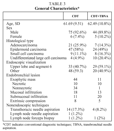
The cases of 140 patients diagnosed with bronchogenic carcinoma from January 1999 through December 2001 were analyzed retrospectively. Fiberoptic bronchoscopy was performed on all patients, with visible endobronchial lesion defined as exophytic mass, mucosal infiltration (consisting of abnormalities or granuloma in the bronchial wall with friable mucosa), submucosal infiltration (with thickening or loss of longitudinal mucosal folds) and extrinsic compression (swelling of lung walls or carinal widening).
Procedure
The examinations were carried out by three different specialists and bronchial washings, bronchial brushings, and forceps biopsy samples were essential requisites. When the bronchoscopist considered TBNA was indicated, it was carried out prior to other techniques. For the patient to be included in our study at least 2 bronchial brushings, 3 forceps biopsies, and 2 TBNAs were required.
Cytological analysis was considered positive only when a sufficient number of definitely malignant cells was observed. Cellular atypia and abnormal cells highly suggestive of malignancy were considered negative. Samples were immediately fixed in 95% proof alcohol; all samples were assessed by the same cytologist, who was blinded to the histological techniques used. In all TBNA cases, 22-gauge needles (MW-222; Mill-Rose Lab, Mentor, OH, USA) were used; disposable catheters 1.7 mm in diameter (1601 Boston Scientific, Watertown, MA, USA) were used for bronchial brushings. The choice of forceps for biopsy was left to the bronchoscopist in charge.
Exclusion Criteria
Patients were withdrawn when thoracotomy was required to classify the neoplasm, or when the cytology samples were considered inadequate.
Variables
First, the diagnostic yield for CDT was compared to the yield for CDT+TBNA. The diagnostic positivity by bronchoscopy was determined in both groups as a function of the visualized lesion.
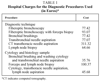
Second, the cost of diagnosis was calculated in euros using the figures provided by the billing department at our hospital. The cost per diagnosis was the sum of costs needed to reach a diagnosis including endoscopic and other procedures such as transthoracic needle aspiration, and lymph node biopsy. The charges for diagnostic procedures used are listed in Table 1. The costs of analyzing samples after the various endoscopic diagnostic procedures were included for both CDT and CDT+TBNA cases.
Statistical Analysis
The results of data analysis of quantitative variables are expressed as means (SD). Percentages were used for the qualitative variables. Percentages were compared using χ² tests. Independent sample means were compared with Student t tests.
Results
Of the 140 patients enrolled in the study, 10 were excluded on the following grounds: 2 because they had undergone thoracotomy and 8 because their samples were considered inadequate. Therefore, the study population was made up of 130 patients: 120 men (91.5%), with a mean age of 62.02 (9.90) years. During the examination, the following lesions were observed: exophytic mass in 55 patients, mucosal infiltration in 31, extrinsic compression in 13, and submucosal infiltration in 31. The histological classification was epidermoid carcinoma for 71 patients (54.6%), adenocarcinoma for 28 (21.5%), microcytic carcinoma in 17 (13.1%) and undifferentiated large cell carcinoma in 14 (10.8%). TNBA was performed on 49 patients and its diagnostic yield of 85.7% was higher than that of any other technique (Table 2). No serious complications related to the procedure were observed except on 2 occasions in which moderate bleeding occurred. Bleeding was controlled by conventional endoscopic means. Figure 1 shows the number of TBNAs performed by each practitioner as well as the diagnostic yield obtained.
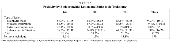
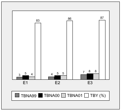
Figure 1. Transbronchial needle aspirations and yields by endoscopist. E1 indicates endoscopist 1; E2, endoscopist 2; E3, endoscopist 3; TBNA99, transbronchial needle aspirations in 1999; TBNA00, transbronchial needle aspirations in 2000; TBNA01, transbronchial needle aspirations in 2001; TBY, transbronchial needle aspiration yield.
Diagnostic Yield for Fiberoptic Bronchoscopy
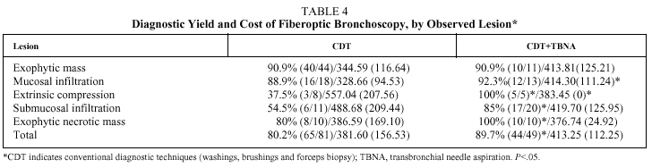
CDT led to cytohistological diagnosis in 80.2% of cases and CDT+TBNA gave positive results in 89.7% (P=.01). The gain in diagnostic yield continued to be significant for the following lesions: extrinsic compression (CDT: 37.5%; CDT+TBNA: 100%; P=.01), submucosal infiltration (CDT: 54.5%; CDT+TBNA: 85%; P=.03), and exophytic mass with surface necrosis (CDT: 80%: CDT+TBNB:100%; P=.01). Table 4 shows the diagnostic yield by the type of endobronchial lesion and presence of necrosis.


Study of Costs
The mean (SD) cost per disease diagnosed was € 393.53 (€ 142.04): € 381.60 (€ 156.53) with CDT and € 431.25 (€ 112.91) in CDT+TBNA.
Table 4 shows that the addition of TBNA lowered costs when submucosal disease (CDT: € 488.68 [€ 209.44]; CDT+TBNB: € 419.70 [€ 125.95]), exophytic mass with necrosis (CDT: € 386.59 [€ 169.10]; CDT+TBNB: € 376.74 [€ 24.92]), and extrinsic compression (CDT: € 557.04 [207.56]; CDT+TBNB: € 383 [€ 0]; P=.02) were present.
Discussion
For the staging of lung carcinoma TBNA has been widely studied and is recommended as part of standard medical practice in various scientific associations' guidelines, the same cannot be said of its use in the diagnosis of an endoscopically visible lesion. Few authors have evaluated its utility in this respect.
TBNA diagnosed malignancy in 85.7% of the patients analyzed retrospectively in the present study and obtained the highest yield of all techniques, proving better than forceps biopsy for all endobronchial anomalies. It was also the only procedure able to establish the diagnosis in 17% of cases. The addition of TBNA to conventional cytology and histology techniques significantly increased the diagnostic yield of the endoscopic exploration by 9.5%, to reach a yield of 89.7%. Increased yield was observed for exophytic mass lesions with surface necrosis, submucosal disease, and extrinsic compression.
Other authors have reported similar results. For example, in a prospective analysis by Govert et al,6 TBNA showed a sensitivity of 79% for classifying malignancy; TBNA plus forceps biopsy and bronchial brushings positivity increased positivity to 95%, although greatest usefulness of TBNA was observed in extrinsic compression and submucosal infiltration. In a similar study by Dasgupta et al7 TBNA obtained an overall yield of 85%; TBNA plus forceps biopsy and brushing increased the yield to 96% in cases of exophytic mass lesion, submucosal disease, and extrinsic compression. Similarly, diagnostic yields ranging from 82% to 97% have been reported for submucosal infiltration.8,9
In short, the usefulness of TBNA seems beyond question. Nonetheless, it is important to remember that the aim of a new technique is to increase diagnostic yield and reduce the cost10 of diagnosing patients with lung carcinoma. According to Govert el al,5 a cytology diagnosis that increases the yield of endoscopy by 6% is cost effective, and so the regular use of endoscopy seems advisable based on our results and the literature cited. We should also remember that the endoscopist´s aim is to reduce the number of explorations that fail to provide a diagnosis and to avoid the use of additional techniques.11 Taking all these points into consideration, we analyzed the cost of both endoscopic and nonendoscopic techniques needed for cytohistological typing. Table 4 shows that TBNA combined with other techniques reduces the number of endoscopies failing to provide diagnoses, mainly when the lesion visualized is submucosal infiltration, exophytic mass with necrosis, or extrinsic compression. This is reflected in lower costs, although only in the case of extrinsic compression is the saving significant.
Our deduction is based on the assumptions outlined, TBNA meets the necessary requirements for regular use in the diagnosis of bronchogenic carcinoma with visible endoscopic lesions.
Our study may suffer from a certain sampling bias given the possibility inherent to its retrospective design that there was a certain degree of variability in the criteria the 3 endoscopists used when describing and interpreting the lesions visualized. Variation in the yield of bronchoscopy might also have been present. Similarly, although the cytology samples were always analyzed by the same pathologist, 2 different groups handled the histology specimens. Nevertheless, in our judgment, the impact on the study results of having different groups was minor, since our endoscopists and pathologists have had solid experience that allowed them to define the anomaly observed in similar ways, with no significant differences among them in diagnostic yield. Furthermore, all the data were collected by the same person using a standard protocol and for the inclusion of a case in the study we required a minimum number of samples to have been taken, following previously established guidelines.
In spite of these limitations, we believe that our results are valid and they acquire particular importance for 2 reasons: a) given the importance of factors that have nothing to do with endoscopic exploration, such as an effect of the observer12 or pathologist in charge, any procedure which helps to optimize the yield of fiberoptic bronchoscopy would be of great assistance, and b) even though the role of TBNA is acknowledged by several expert committees13 to be quite important, it remains an underutilized technique probably due to a lack of awareness of its advantages, as shown by surveys.14,15 Further studies that demonstrate the safety and cost effectiveness of the technique will undoubtedly be of great assistance in overcoming this obstacle.
We conclude that TBNA is a technique that substantially increases the yield of endoscopic exploration for cases of endobronchial lesions suggestive of neoplasia, with no negative impact on the cost of the diagnostic process, when the lesion corresponds to submucosal disease, exophytic mass with necrosis, or extrinsic compression.
Correspondence: Dr. J.A. Gullón Blanco.
Pérez Galdós, 11, 5.º dcha. 38002 Santa Cruz de Tenerife. España.
E-mail: jose993@separ.es
Manuscript received February 11, 2003.
Accepted for publication May 27, 2003.


