
To the editor: Soft tissue sarcomas are neoplasms which establish themselves on structures of mesodermic origin. The most common histological variant in adults is the malignant fibrous histiocytoma (MFH), though this rarely occurs in the lung.1 Furthermore, before labeling such a histiocytoma as being of pulmonary origin, it is important to rule out the possibility of a primary site elsewhere, given that such tumors are more often the result of metastasis. We report a case of this unusual pulmonary disease.
A clinically asymptomatic 77-year-old man with a history of cutaneous melanoma of superficial extension (Breslow depth 2.3 mm) extirpated two years previously was seen in a routine x-ray to have a nodule at the base of the right lung. Physical examination revealed nothing abnormal apart from hepatomegaly of 4 cm due to alcohol-related cirrhosis. All tests, including tumor and coagulation markers, provided normal results, with the exception of an increase in bilirubin (2 mg/dL) and transaminases because of the liver disease. In the lateral segment of the inferior lobe of the right lung, computed axial tomography revealed a nodular, lobulate image 1.5 cm in diameter which had not appeared in tests the previous year. The results of fine needle aspiration did not conclusively rule out malignancy. On the suspicion that the object represented a single metastasis of the melanoma, the patient underwent a serratus-sparing right posterolateral thoracotomy, and a 2-cm nodule found in the inferior right lobe was extirpated by means of an inferior lobectomy. A histological exam revealed a mesenchymal neoformation that had infiltrated the pulmonary parenchyma, with areas of fascicular-storiform pattern intermingled with areas of hyaline fibrosis. Fusiform cells with enlarged nuclei of atypical size and atypical multinucleate giant cells with nucleoli and frequent mitoses, some cells showing atypical morphology, were observed. In other areas a nonspecific lymphoplasmacytic inflammatory component was evident. In immunohistochemical testing, the neoplastic cells showed positive for vimentin and CD-68 (Figure), but negative for S-100 protein, actin, HMB-45, and cytokeratin, which are specific markers for melanoma. The postoperative period was free of complications. Two years after the intervention, the patient continues to be asymptomatic and free of lung disease.

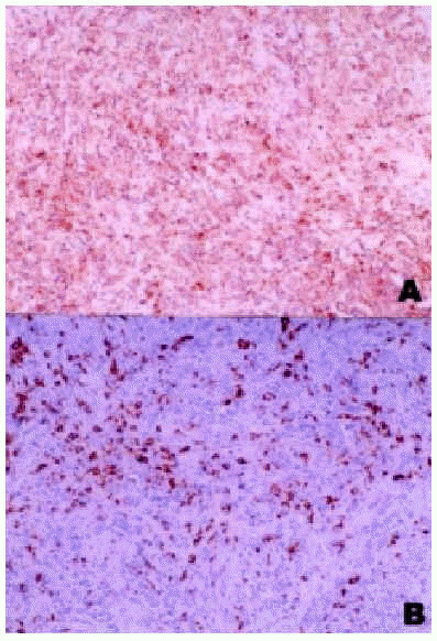
Figure. Histology, immunohistological assay. A: expression of vimentin by neoplastic cells (vimentin, x250); B: expression of CD-68 by neoplastic cells (CD-68, x250).
Pulmonary MFH tends to occur in patients in their 60s, with no differences in incidence between the sexes. Clinical signs and symptoms are typically chest pain and dyspnea, often accompanied by cough, weight loss, hemoptysis, and fatigue. Asymptomatic cases such as the one described are rare.1 Their prognosis is very poor and depends primarily on early diagnosis, given that the cases involving long survival have been described at their initial stages when resective surgery was possible.1-4 It is essential to rule out both remote metastasis of MFH and sarcomatoid carcinoma, malignant desmoplastic mesothelioma, and all other types of pulmonary sarcoma.1-4
Radiographically, MFHs tend to be solitary, noncavitated and peripheral, located most often in the middle or inferior lobe, as in this case, and they frequently affect the pleura and are associated with pleural effusion.1-4 The typical tomographic image is of a large, well-circumscribed tumor, neither cavitated nor calcified, located peripherally and without hilar or mediastinal adenopathy.1 Bronchoscopy and fine needle aspiration are of limited value in the differential diagnosis,3 making it often necessary to perform a thoracotomy in order to reach the definitive diagnosis, as was true in this case. Microscopic inspection shows a malignant neoplasm of spicular cells. Unfortunately there exist no specific immunohistological markers, though similar sarcomas can be ruled out by means of specific immunohistochemical assays. Histological variants are the storiform, pleomorphic, myxoid, inflammatory, giant cell, and angiomatoid variants, with the majority of pulmonary MFHs being of a mixed storiform-pleomorphic variety.
Resective surgery is the standard, essential treatment. It has been noted that if treatment is limited to biopsy or partial resection, the survival rate averages between 9-10 months,1 whereas if the surgery is intended to be curative, the disease-free survival improves, with 20% alive at 5 years.1-4 Magne et al5 have studied survival rates for cases of primary pulmonary sarcoma and observed that rates are significantly higher when total resection is performed compared with partial surgery (47 vs 6 months). Furthermore, survival longer than 5 years has been obtained with complete resections of recurrences, early diagnosis being essential in these instances; hence the importance of periodic check-ups. Most authors have not been able to demonstrate the usefulness of radiotherapy in treatment, though Lee et al2 achieved extended survival by applying 5400 cGy prior to surgery. Systemic chemotherapy is used for metastatic cases but has not succeeded in improving survival.1-4
Systemic metastases, primarily cerebral, are common, which may be due to the high incidence of vascular invasion at the time of diagnosis.1 The prognostic factors involved in pulmonary location are not well understood, although, in general terms, total resection of the macroscopic disease, age above 50 years, the tumor grade, the histological variant, the size of the tumor, and the number of distant metastases at the time of diagnosis have been described as prognostic factors in MFH.6 In the pulmonary cases that have been described, nearly all patients with long survival have been negative for node involvement, as was the case with our own patient1-3; only Yousen and Hochholzer4 have described a patient who survived for 36 months with mediastinal adenopathy. No clear relation between survival and pulmonary histology has been established, possibly because of the predominance of the mixed storiform-pleomorphic subtype.


