
Hydrocephalus is the abnormal dilation of the ventricular system of the brain due to the accumulation of cerebrospinal fluid (CSF). There are various methods for shunting CSF from the central nervous system to other cavities with absorptive capacity, the most widely used of these being the ventriculo-peritoneal (VP) shunt. Under certain circumstances, this type of shunt placement is unadvisable or contraindicated: infections, previous surgeries that could favor the development of bridles, thrombosis or obliteration of the drainage system. In such cases, ventriculo-pleural (VPL) shunts are a simple, safe alternative. This technique, however, is not free of complications.
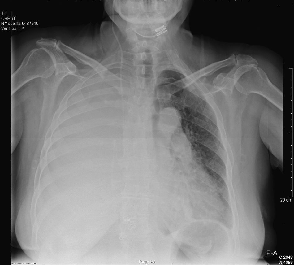
We present the case of a 59-year-old woman with a history of normal-pressure hydrocephalus requiring treatment with VP shunting. Eight years later, the patient underwent abdominal surgery due to acute perforated diverticulitis. The post-surgery evolution was torpid, with episodes of repetitive meningitis and a nasal CSF fistula. Given the said complications and the previous abdominal surgery, a decision is made to place a programmable VPL shunt. After a few months, the patient was admitted to the ICU due to progressive respiratory failure, even at rest, with criteria for severity. Upon auscultation, the patient presented diminished vesicular murmur in the right hemithorax, and chest radiography revealed a massive right pleural effusion (Fig. 1). Thoracocentesis was performed and an intrathoracic drain tube was inserted. The liquid presented biochemical characteristics compatible with hydrothorax. After increasing the pressure of the valve aperture and the evacuation of the effusion, the later evolution was satisfactory.
Since the description of VPL shunting by Heile in 1914, this technique has been a useful alternative to VP shunts, which today is still the method of choice.1 CSF can be directed towards different cavities, such as the peritoneal, atrial and pleural. Important groups of complications have been reported: mechanical (obstruction of the catheter, migration or rupture), functional (due to poor CSF absorption) and infectious (with an incidence of 8%–12% in the first 6 months). Ventriculo-atrial shunts have fallen into disuse because of thromboembolic complications (pulmonary, intracardiac, vena cava) and infections (thrombophlebitis, septic emboli, nephritis). Thus, the use of VPL shunts is a simple, useful option, although it is not complication-free. Initially, its use was conceived as a temporary derivation while infections related with VP shunts were resolved. In 1954, Ransohoff reported a series of 6 patients with hydrocephalus secondary to tumor pathology that were successfully treated with VPL shunting, giving evidence of the good absorptive capacity of the pleura, but the long-term results were not satisfactory.2 Later, in 1988, Jones published a series of 29 children treated with VPL shunts, in only 7 of whom they were maintained functional for more than a year.3 Megison and Benzell affirmed that children and adults with lung disease should not be candidates for this technique due to the lower absorptive capacity.4 The appearance of pressure valves with regulatable aperture and anti-siphon mechanisms has allowed for better control of CSF drainage, reducing the incidence of symptomatic pleural effusion. Nevertheless, the introduction of these valves can cause, inversely, effects related with the limited drainage of CSF due to the narrow control margins of the intracranial pressure.
Despite all the advances made in its management, the mechanisms by which CSF accumulates pathologically after VPL shunt placement are still unknown. It has been postulated that there are irritative or inflammatory mechanisms related with the catheter itself (generating a greater presence of lymphocytes in the pleural liquid). This mechanism generates an increase in the production of pleural liquid with alteration of the lymphatic flow, as well as a reduction of the pleural absorptive surface. This latter point is especially true in children, seniors and patients with lung pathologies. The establishment of this phenomenon can be gradual, which would explain the evolution of the disease observed in our patient.
Despite the lack of prospective studies evaluating the effectiveness of VPL shunting, we can conclude that it is an effective alternative for long-term CSF drainage when there are contraindications for VP shunt placement.5 We should, therefore, become familiar with this technique and keep in mind its potential complications in order to detect these early on.
Please cite this article as: Gascón-Sánchez V, et al. Insuficiencia respiratoria grave secundaria a drenaje ventriculopleural. Arch Bronconeumol. 2011;47:477-8.


