array:19 [
  "pii" => "13050406"
  "issn" => "15792129"
  "estado" => "S300"
  "fechaPublicacion" => "2003-08-01"
  "documento" => "article"
  "crossmark" => 0
  "subdocumento" => "fla"
  "cita" => "Arch Bronconeumol. 2003;39:361-6"
  "abierto" => array:3 [
    "ES" => false
    "ES2" => false
    "LATM" => false
  ]
  "gratuito" => false
  "lecturas" => array:2 [
    "total" => 29204
    "formatos" => array:3 [
      "EPUB" => 221
      "HTML" => 26795
      "PDF" => 2188
    ]
  ]
  "itemSiguiente" => array:15 [
    "pii" => "13050407"
    "issn" => "15792129"
    "estado" => "S300"
    "fechaPublicacion" => "2003-08-01"
    "documento" => "article"
    "crossmark" => 0
    "subdocumento" => "fla"
    "cita" => "Arch Bronconeumol. 2003;39:367-9"
    "abierto" => array:3 [
      "ES" => false
      "ES2" => false
      "LATM" => false
    ]
    "gratuito" => false
    "lecturas" => array:2 [
      "total" => 3078
      "formatos" => array:3 [
        "EPUB" => 139
        "HTML" => 2212
        "PDF" => 727
      ]
    ]
    "en" => array:9 [
      "idiomaDefecto" => true
      "titulo" => "Don Antonio Corbella y Fondebila's Treatise on Chest Diseases"
      "tienePdf" => "en"
      "tieneTextoCompleto" => "en"
      "paginas" => array:1 [
        0 => array:2 [
          "paginaInicial" => "367"
          "paginaFinal" => "369"
        ]
      ]
      "titulosAlternativos" => array:1 [
        "es" => array:1 [
          "titulo" => "El tratado de las enfermedades del pecho de D. Antonio Corbella y Fondebila"
        ]
      ]
      "contieneTextoCompleto" => array:1 [
        "en" => true
      ]
      "contienePdf" => array:1 [
        "en" => true
      ]
      "autores" => array:1 [
        0 => array:2 [
          "autoresLista" => "J Sauret Valet"
          "autores" => array:1 [
            0 => array:2 [
              "Iniciales" => "J"
              "apellidos" => "Sauret Valet"
            ]
          ]
        ]
      ]
    ]
    "idiomaDefecto" => "en"
    "EPUB" => "https://multimedia.elsevier.es/PublicationsMultimediaV1/item/epub/13050407?idApp=UINPBA00003Z"
    "url" => "/15792129/0000003900000008/v0_201307090911/13050407/v0_201307090912/en/main.assets"
  ]
  "itemAnterior" => array:15 [
    "pii" => "13050405"
    "issn" => "15792129"
    "estado" => "S300"
    "fechaPublicacion" => "2003-08-01"
    "documento" => "article"
    "crossmark" => 0
    "subdocumento" => "fla"
    "cita" => "Arch Bronconeumol. 2003;39:353-60"
    "abierto" => array:3 [
      "ES" => false
      "ES2" => false
      "LATM" => false
    ]
    "gratuito" => false
    "lecturas" => array:2 [
      "total" => 3630
      "formatos" => array:3 [
        "EPUB" => 143
        "HTML" => 2776
        "PDF" => 711
      ]
    ]
    "en" => array:11 [
      "idiomaDefecto" => true
      "titulo" => "Prognostic Factors of Morbidity and Mortality in the Early Postoperative Period Following Lung Transplantation"
      "tienePdf" => "en"
      "tieneTextoCompleto" => "en"
      "tieneResumen" => array:2 [
        0 => "en"
        1 => "es"
      ]
      "paginas" => array:1 [
        0 => array:2 [
          "paginaInicial" => "353"
          "paginaFinal" => "360"
        ]
      ]
      "titulosAlternativos" => array:1 [
        "es" => array:1 [
          "titulo" => "Factores pronósticos de morbimortalidad en el postoperatorio inmediato del trasplante pulmonar"
        ]
      ]
      "contieneResumen" => array:2 [
        "en" => true
        "es" => true
      ]
      "contieneTextoCompleto" => array:1 [
        "en" => true
      ]
      "contienePdf" => array:1 [
        "en" => true
      ]
      "autores" => array:1 [
        0 => array:2 [
          "autoresLista" => "FJ Gómez, A Planas, P Ussetti, JJ Tejada, A Varela"
          "autores" => array:5 [
            0 => array:2 [
              "Iniciales" => "FJ"
              "apellidos" => "Gómez"
            ]
            1 => array:2 [
              "Iniciales" => "A"
              "apellidos" => "Planas"
            ]
            2 => array:2 [
              "Iniciales" => "P"
              "apellidos" => "Ussetti"
            ]
            3 => array:2 [
              "Iniciales" => "JJ"
              "apellidos" => "Tejada"
            ]
            4 => array:2 [
              "Iniciales" => "A"
              "apellidos" => "Varela"
            ]
          ]
        ]
      ]
    ]
    "idiomaDefecto" => "en"
    "EPUB" => "https://multimedia.elsevier.es/PublicationsMultimediaV1/item/epub/13050405?idApp=UINPBA00003Z"
    "url" => "/15792129/0000003900000008/v0_201307090911/13050405/v0_201307090912/en/main.assets"
  ]
  "en" => array:11 [
    "idiomaDefecto" => true
    "titulo" => "Respiratory Muscle Disorders in Chest Wall Diseases"
    "tieneTextoCompleto" => true
    "paginas" => array:1 [
      0 => array:2 [
        "paginaInicial" => "361"
        "paginaFinal" => "366"
      ]
    ]
    "autores" => array:1 [
      0 => array:3 [
        "autoresLista" => "A Casas, J Pavía, D Maldonado"
        "autores" => array:3 [
          0 => array:3 [
            "Iniciales" => "A"
            "apellidos" => "Casas"
            "referencia" => array:1 [
              0 => array:2 [
                "etiqueta" => "<span class="elsevierStyleSup">a</span>"
                "identificador" => "affa"
              ]
            ]
          ]
          1 => array:3 [
            "Iniciales" => "J"
            "apellidos" => "Pav&#237;a"
            "referencia" => array:1 [
              0 => array:2 [
                "etiqueta" => "<span class="elsevierStyleSup">a</span>"
                "identificador" => "affa"
              ]
            ]
          ]
          2 => array:3 [
            "Iniciales" => "D"
            "apellidos" => "Maldonado"
            "referencia" => array:1 [
              0 => array:2 [
                "etiqueta" => "<span class="elsevierStyleSup">a</span>"
                "identificador" => "affa"
              ]
            ]
          ]
        ]
        "afiliaciones" => array:1 [
          0 => array:3 [
            "entidad" => "Fundación Neumológica Colombiana. Bogotá. Colombia."
            "etiqueta" => "<span class="elsevierStyleSup">a</span>"
            "identificador" => "affa"
          ]
        ]
      ]
    ]
    "titulosAlternativos" => array:1 [
      "es" => array:1 [
        "titulo" => "Trastornos de los m&#250;sculos respiratorios en las enfermedades de la pared del t&#243;rax"
      ]
    ]
    "textoCompleto" => "<p class="elsevierStylePara"><span class="elsevierStyleBold">Introduction</span></p><p class="elsevierStylePara"> Diseases that constrict the skeletal structure of the chest wall and the spine with its articulations may interfere with the functional capacity of the diaphragmatic pump&#44; facilitating the development of respiratory insufficiency and failure&#46;</p><p class="elsevierStylePara">The present article discusses the response of the respiratory muscles to mechanical overload&#46; Following a brief examination of the force these muscles are required to exert in normal conditions&#44; we review the pathophysiology of some characteristic alterations brought about by <span class="elsevierStyleItalic">a&#41;</span> chest wall diseases such as kyphoscoliosis&#59; <span class="elsevierStyleItalic"> b&#41;</span> other diseases involving the bony structure of the chest wall such as ankylosing spondylitis and pectus excavatum<span class="elsevierStyleItalic">&#59;</span> and <span class="elsevierStyleItalic">c&#41;</span> extreme obesity&#46;</p><p class="elsevierStylePara"> Systemic disorders of a neuromuscular type will be dealt with in a different chapter of the series&#46;</p><p class="elsevierStylePara"><span class="elsevierStyleBold">The structure and function of the chest wall</span></p><p class="elsevierStylePara">The chest wall is configured by the rib cage&#44; the spinal column&#44; and the diaphragm&#46; The movements of the chest wall depend on a complex interaction among these components&#46; The diaphragm forms a domelike muscular-tendinous partition which is displaced some 9&#46;5 cm on average during tidal volume generation and which affects the chest wall by changes caused in abdominal and pleural pressures&#46; During normal respiration&#44; a contact point or &#34;zone of apposition&#34; is established between the lateral part of the base of this diaphragmatic dome and the internal surface of the lower ribs&#46; During inspiration&#44; the fibers of the diaphragm shorten and thus impart a caudal piston-like movement&#46; This contraction thereby lowers pleural pressure &#40;P<span class="elsevierStyleInf">pl</span>&#41; and raises intra-abdominal pressure&#46; The decrease in P<span class="elsevierStyleInf">pl</span> determines the expansion of the lungs and the increase in abdominal pressure&#44; which is transmitted to the lower part of the chest wall via the apposition zone&#44; which in turn makes the lower ribs expand&#46; The movement of the ribs&#44; which depends on their insertions into the sternum and into the vertebral column&#44; is determined by the contraction of the parasternal muscles and the scalene muscles&#44; which insert into the 1st to 6th ribs&#44; and also by the costal portion of the diaphragm&#44; which inserts into the sternum and into the 7th to 12th ribs&#46; The muscles of the abdominal wall are important in expiration and their contraction has a significant effect on the lung volume&#46; Electromyography &#40;EMG&#41; of the inspiratory muscles at rest shows that the inspiratory intercostals&#44; the parasternals&#44; and the scalenes act phasically in inspiration and are thus now considered accessory muscles&#46; Deep breathing or exhausting exercise activates muscles such as the sternocleidomastoids&#44; which should be considered the true accessory muscles&#46;</p><p class="elsevierStylePara"><span class="elsevierStyleItalic">The mechanics of inspiration and expiration</span></p><p class="elsevierStylePara">The usual end-expiratory lung volume in normal subjects &#40;functional residual capacity or FRC&#41; represents the perfect balance point or rest point between the elastic recoil pressures of the chest wall &#40;P<span class="elsevierStyleInf">W</span>&#41; and the lung wall &#40;P<span class="elsevierStyleInf">L</span>&#41;&#46; The pressure applied by the inspiratory muscles has to overcome three types of forces or resistances that oppose a change in volume&#58; <span class="elsevierStyleItalic">a&#41;</span> elastic forces&#44; which are related to volume change&#59; <span class="elsevierStyleItalic">b&#41;</span> resistive forces&#44; which are related to airflow &#40;volume change per unit of time&#41;&#59; and <span class="elsevierStyleItalic">c&#41;</span> inertia&#44; which is related to the initial force required to move tissues and air and which is not considered significant under normal conditions&#46; The brief description presented here of the elastic properties of the lung and chest wall and of airway resistance should facilitate analysis of their complex interaction&#46;</p><p class="elsevierStylePara">In normal conditions &#40;Figure 1&#41;&#44; at end expiration P<span class="elsevierStyleInf">L</span> is approximately 5 cm H<span class="elsevierStyleInf">2</span>O and P<span class="elsevierStyleInf">W</span> is equal but opposite &#40;&#173;-5 cm H<span class="elsevierStyleInf">2</span>O&#41;&#46; This balance produces a P<span class="elsevierStyleInf">pl</span> of &#173;-5 cm H<span class="elsevierStyleInf">2</span>O and an alveolar pressure &#40;P<span class="elsevierStyleInf">alv</span>&#41; of 0 cm H<span class="elsevierStyleInf">2</span>O&#46; Therefore&#44; there is no airflow&#46; The contraction of the inspiratory muscles will create an expansive force of the chest wall &#40;P<span class="elsevierStyleInf">mus</span>&#41; which is negative in sign and which is transmitted through the pleural space to the alveoli&#44; where the pressure becomes subatmospheric&#44; generating a pressure gradient and thus airflow from the mouth to the alveoli&#46; In a partial inspiration&#44; the negative pressure generated by the inspiratory muscles exceeds P<span class="elsevierStyleInf">L</span> and a subatmospheric P<span class="elsevierStyleInf">alv</span> and inspiratory airflow persist&#46; At the end of an inspiration &#40;at total lung capacity--TLC&#41;&#44; a balance is established between the negative pressure exercised by the inspiratory muscles and the positive pressure exerted by P<span class="elsevierStyleInf">W</span> and P<span class="elsevierStyleInf">L</span>&#46; Once again P<span class="elsevierStyleInf">alv</span> is 0 cm H<span class="elsevierStyleInf">2</span>O and there is no airflow&#46; Expiration begins when contraction of the inspiratory muscles decreases&#59; thus&#44; the elastic recoil pressures P<span class="elsevierStyleInf">W</span> and P<span class="elsevierStyleInf">L</span> predominate&#44; generating a positive P<span class="elsevierStyleInf">alv</span> and expiratory flow which will continue until an equilibrium of P<span class="elsevierStyleInf">W</span> and P<span class="elsevierStyleInf">L</span> is reached&#46; It is now known that there is continuous but decreasing activity of the inspiratory muscles during expiration acting as a brake on expiratory flow&#46;</p><p class="elsevierStylePara"><img src="260v39n08-13050406tab01.gif"></img></p><p class="elsevierStylePara">Fig&#46; 1&#46; Schematic representation of pressure changes in different phases of the respiratory cycle in a healthy subject&#46; P<span class="elsevierStyleInf">W</span> indicates elastic recoil pressure of the chest wall&#59; P<span class="elsevierStyleInf">pl</span>&#44; pleural pressure&#59; P<span class="elsevierStyleInf">L</span>&#44; elastic recoil pressure of the lung&#59; P<span class="elsevierStyleInf">alv</span>&#44; alveolar pressure&#59; P<span class="elsevierStyleInf">mo</span>&#44; mouth pressure&#59; Pmus&#44; muscle contraction pressure&#46; Pressures are expressed in cm H<span class="elsevierStyleInf">2</span>O&#46; The top figure &#40;framed&#41; represents the equivalences&#59; the figures of partial inspiration and expiration represent the same lung volume&#46; See explanation in text&#46; &#40;Adapted from Gibson&#46;<span class="elsevierStyleSup">1</span>&#41;</p><p class="elsevierStylePara">In scoliosis and other chest wall disorders&#44; the elastic properties of the lungs and airways are normal&#59; thus&#44; FRC or end-expiratory pressures are normal&#46; Since there is great reduction of chest wall compliance &#40;chest wall stiffness&#41; in these diseases&#44; greater inspiratory force is required to overcome the reduction without producing very great changes in P<span class="elsevierStyleInf">pl</span>&#46; Figure 2 shows that&#44; assuming that chest wall compliance is half of normal&#44; P<span class="elsevierStyleInf">W</span> at the same tidal volume will increase twofold over the normal level &#40;eg&#44; from &#173;-5 to &#43;3 cm H<span class="elsevierStyleInf">2</span>O&#44; as opposed to &#173;-5 to -&#173;1 cm H<span class="elsevierStyleInf">2</span>O in healthy subjects&#41; between the end of expiration and the end of inspiration&#46; Therefore&#44; greater inspiratory force is required to overcome chest wall stiffness&#46;</p><p class="elsevierStylePara"><img src="260v39n08-13050406tab02.gif"></img></p><p class="elsevierStylePara">Fig&#46; 2&#46; Schematic representation of pressure changes in different phases of the respiratory cycle in a subject with a chest wall disease or chest wall stiffness&#46; See Figure 1 for equivalences&#46; FRC indicates functional residual capacity&#59; TLC&#44; total lung capacity&#46; The figures of partial inspiration and expiration represent the same lung volume&#46; See explanation in text&#46; &#40;Adapted from Gibson&#46;<span class="elsevierStyleSup">1</span>&#41;</p><p class="elsevierStylePara"><span class="elsevierStyleItalic">Lung and chest wall distensibility</span></p><p class="elsevierStylePara">The lungs and the chest wall are elastic structures that tend to return to their initial shape after having been deformed by an external force&#46; The elastic recoil pressure is that which opposes their stretch or distension&#46; The system elasticity may be expressed as compliance and defined as volume change per unit pressure change &#40;&#916;V&#47;&#916;P&#41;&#46;</p><p class="elsevierStylePara">In the healthy subject a Rahn diagram &#40;Figure 3a&#41; illustrates that TLC is determined by the balance between the pressures generated by the respiratory muscles and the elastic recoil of the respiratory system&#46; In a deep breath with an open glottis&#44; P<span class="elsevierStyleInf">alv</span> is zero &#40;transrespiratory system pressure&#44; or P<span class="elsevierStyleInf">RS</span>&#44; equals -&#173;P<span class="elsevierStyleInf">mus</span>&#41;&#46; In patients with chest wall diseases&#44; which are characterized by increased chest wall stiffness &#40;Figure 3b&#41;&#44; P<span class="elsevierStyleInf">W</span> and thus P<span class="elsevierStyleInf">RS</span> will increase&#44; meaning that balance &#40;P<span class="elsevierStyleInf">RS</span> &#61; &#173;-P<span class="elsevierStyleInf">mus</span>&#41; will occur at a much lower volume than that observed in healthy persons&#44; such that the pressure-volume curve is flatter&#46; Because the volume is reduced&#44; the maximum P<span class="elsevierStyleInf">L</span> is lower than normal&#46;</p><p class="elsevierStylePara"><img src="260v39n08-13050406tab03.gif"></img></p><p class="elsevierStylePara">Fig&#46; 3&#46; Pressure-volume curve determinants in a healthy subject &#40;a&#41; and in a subject with chest wall disease or chest wall stiffness &#40;b&#41;&#46; P<span class="elsevierStyleInf">W</span> indicates elastic recoil pressure of the chest wall&#59; P<span class="elsevierStyleInf">RS</span>&#44; transrespiratory system pressure&#59; P<span class="elsevierStyleInf">L</span>&#44; elastic recoil pressure of the lung&#59; P<span class="elsevierStyleInf">mus</span>&#44; muscle contraction pressure&#59; FRC&#44; functional residual capacity&#59; TLC&#44; total lung capacity&#46; Pressures are expressed as cm H<span class="elsevierStyleInf">2</span>O&#46; See explanation in text&#46;</p><p class="elsevierStylePara"><span class="elsevierStyleItalic">Evaluation respiratory muscle functions</span></p><p class="elsevierStylePara">The mechanical properties and function of the respiratory muscles can be evaluated by measuring lung volumes&#44; chest wall motion&#44; and pressures at different locations of the chest wall&#46; Respiratory muscle activity can also be characterized by EMG&#46;</p><p class="elsevierStylePara">In the respiratory system&#44; force is measured as pressures&#44; and muscle contraction is measured as volume changes or displacement of chest wall structures&#46; To characterize the respiratory muscles quantitatively&#44; we can use measurements of volume&#44; displacement&#44; pressures&#44; and the relationships between changes in these variables over time&#46; Thoracic geometry has an important role to play in efficiently converting force into pressure&#46; These pressures also depend on the mechanical characteristics of the thoracic cage and the abdominal wall&#44; with which respiratory muscles interact&#46; A stiff chest wall resists the distortion generated by muscle action and thus permits the diaphragm to create greater pressure for a given volume change&#46; Therefore&#44; the pressures should be seen only as indices of the overall work of the respiratory muscles and not as a direct measure of their contraction&#46; The global evaluation of the respiratory muscles has been dealt with in other chapters of this series&#59; thus&#44; we need not enter into a discussion of this particular aspect at this time&#46;<span class="elsevierStyleSup">2</span></p><p class="elsevierStylePara"><span class="elsevierStyleBold">Kyphoscoliosis</span></p><p class="elsevierStylePara"> Kyphoscoliosis is a disease of the vertebral column and its articulations&#44; characterized by the simultaneous presence of scoliosis &#40;lateral curvature of the spine&#41; and kyphosis &#40;anteroposterior spinal curvature&#41;&#46; Signs of the latter may be destruction of vertebral bodies&#44; bone tuberculosis&#44; osteoporosis&#44; etc&#46; Primary or idiopathic kyphoscoliosis is a hereditary disease characterized by defects in the development of the vertebrae or by connective tissue disease&#46; It accounts for 80&#37; of cases and is more frequent in women&#46; Secondary kyphoscoliosis generally originates in childhood neuromuscular diseases such as poliomyelitis or muscular dystrophies&#46;<span class="elsevierStyleSup">3</span></p><p class="elsevierStylePara"> During the course of kyphoscoliosis&#44; the initial displacement of the spine &#40;&#34;primary curvature&#34;&#41; involves two structural changes&#58; on the one hand&#44; a secondary curvature that develops to counterbalance the primary curve and&#44; on the other hand&#44; a rotation of the column on its longitudinal axis&#44; displacing the ribs of the convex side in a posterior and outward direction to form a &#34;hump&#34; shape while the ribs on the concave side are constricted&#46; The intensity of the deformity can be quantified by measuring the angles of the primary convexity&#46; With angles greater than 100&#186;&#44; the deformity is considered severe and the risk of respiratory failure&#44; alveolar hypoventilation&#44; and cor pulmonale is very high&#46;<span class="elsevierStyleSup">4</span></p><p class="elsevierStylePara"> Kyphoscoliosis is expressed in functional changes with decreases in TLC&#44; vital capacity &#40;VC&#41;&#44; and FRC but without changes in residual volume &#40;RV&#41;&#46;<span class="elsevierStyleSup">5&#44;6</span> The severity of compression is directly related to the degree of kyphoscoliotic angulation&#46;<span class="elsevierStyleSup">7-9</span> Diminished expiratory flows are due to the restrictive process <span class="elsevierStyleItalic"> per se&#44;</span> not to intrinsic airway disease&#46;<span class="elsevierStyleSup">10</span></p><p class="elsevierStylePara">In advanced kyphoscoliosis&#44; inspiratory muscle strength is significantly reduced and maximum transdiaphragmatic &#40;Pdi<span class="elsevierStyleInf">max</span>&#41; and inspiratory &#40;PI<span class="elsevierStyleInf">max</span>&#41; pressures are much reduced&#46;<span class="elsevierStyleSup">6</span> In such patients&#44; high positive gastric pressures are generated during respiration&#44; indicating that diaphragm contraction is effective with very negative esophageal pressures&#46; This suggests active recruitment of the intercostal muscles and the inspiratory accessory muscles&#46; In studies of kyphoscoliosis patients&#44; an inverse correlation has been observed between PaCO<span class="elsevierStyleInf">2</span> and PI<span class="elsevierStyleInf">max&#44;</span>&#44; possibly indicating that inspiratory muscle restriction plays a predominant role in the development of respiratory failure in these patients&#46;</p><p class="elsevierStylePara">In patients with chronic obstructive pulmonary disease &#40;COPD&#41; and other similar respiratory diseases&#44; increased nerve activity has been observed in the inspiratory muscles of the rib cage and the diaphragm&#46; Such observations suggest that a typical response to mechanical overload is involved&#46;<span class="elsevierStyleSup">11</span> Consistent with this hypothesis&#44; Estenne et al<span class="elsevierStyleSup">12</span> observed a PI<span class="elsevierStyleInf">max</span>&#44; a sharp decrease in dynamic lung compliance &#40;0&#46;04&#177;0&#46;03 L&#47;cm H<span class="elsevierStyleInf">2</span>O&#41; and an increase in respiratory muscle recruitment during respiration in 8 kyphoscoliotic patients with a severe restrictive pattern &#40;TLC 44&#37;&#177;8&#37; of predicted&#44; FRC 60&#37;&#177;11&#37; of predicted&#44; and RV 82&#37;&#177;20&#37; of predicted&#41;&#46; The observations were attributed to an automatic response of central respiratory drive&#46;</p><p class="elsevierStylePara">In summary&#44; decreased respiratory system compliance&#44; conditioned by the stiffness which deformity transmits to the chest wall&#44;<span class="elsevierStyleSup">13</span> is evident in adults with kyphoscoliosis&#46; This brings about a reduction in the TLC and flattens the pressure-volume curve &#40;Figure 3b&#41;&#46; The respiratory load increases so that patients adopt a breathing pattern characterized by small volumes and very high respiratory rates&#44;<span class="elsevierStyleSup">14</span> with increased dead space&#44; alveolar hypoventilation&#44; and disordered sleep&#44;<span class="elsevierStyleSup">15</span> these changes being very marked at times&#46; Similarly&#44; the greater work of the respiratory muscles stimulates increased oxygen consumption&#44; involving greater risk of respiratory failure due to muscle fatigue&#46;<span class="elsevierStyleSup">16</span></p><p class="elsevierStylePara"> Compliance in children&#44; on the other hand&#44; is preserved regardless of the severity of the deformity&#46;<span class="elsevierStyleSup">17</span></p><p class="elsevierStylePara"><span class="elsevierStyleBold">Ankylosing spondylitis</span></p><p class="elsevierStylePara"> Ankylosing spondylitis is an inflammatory disease of the axial skeleton that mainly affects males&#46; Ninety-five per cent of affected individuals share the genetic marker HLA-B27&#46; Inflammation compromises the ligamentous structures of the spinal column&#44; the sacroiliac articulations&#44; and the ribs&#44; leading to the fibrosis and ossification of these structures and&#44; thus&#44; to an increase in rig cage stiffness due to ankylosis and fusion of the costovertebral and sternoclavicular articulations&#46;</p><p class="elsevierStylePara">In spite of this rib cage stiffness&#44; there is no very marked decrease in the lung volumes&#44; either TLC or FRC&#44;<span class="elsevierStyleSup">18</span> nor is there an observed correlation between the severity of the spinal angulation and the functional parameters indicating restriction&#46; The slight reduction in lung volumes&#44; which is possibly the result of the fusing of the ribs&#44;<span class="elsevierStyleSup">19</span> correlates positively with disease duration and activity&#46;<span class="elsevierStyleSup">20</span> Finally&#44; observed decreases in both PI<span class="elsevierStyleInf">max</span> and maximum expiratory pressure indicate weakness caused by intercostal muscle atrophy plus dysfunction of the diaphragm&#44; whose activity is generally compensatory and preserved&#46;</p><p class="elsevierStylePara"><span class="elsevierStyleBold">Pectus excavatum</span></p><p class="elsevierStylePara"> Pectus excavatum is the most common deformity of the anterior thorax&#46; Consisting of a very notable concavity of the lower third of the sternum&#44; pectus excavatum is a congenital defect which is manifested in different degrees of severity&#46; Thus&#44; it is classified according to the extent of concavity visible in a lateral x-ray of the thorax&#46; It is considered significant when the distance between the surface of the anterior wall of the thorax and the deepest part of the depression is greater than 3 cm&#46;<span class="elsevierStyleSup">21</span> Its etiology is unknown&#44; although a close relation to the Marfan syndrome has been found to exist&#44; suggesting that the connective tissue around the sternum is altered&#46; Lung volumes are at the lower limits of normal or slightly diminished&#44; and compliance is normal unless accompanied by severe kyphoscoliosis&#46;<span class="elsevierStyleSup">22</span></p><p class="elsevierStylePara"><span class="elsevierStyleBold">Obesity</span></p><p class="elsevierStylePara"> When obesity is severe &#40;a body mass index &#91;BMI&#93; greater than 30 kg&#47;m&#178;&#41;&#44; lung function may be compromised&#46; The distribution of obesity can also endanger respiratory function&#44; independently of the total percentage of body fat&#46;<span class="elsevierStyleSup">23</span> However&#44; forced spirometry in patients with uncomplicated obesity is generally normal&#44; although there may be a small decrease in forced VC&#44; which correlates inversely with weight&#46;<span class="elsevierStyleSup">24</span> The effects of uncomplicated obesity are generally reflected more in a significant decrease of the expiratory reserve volume&#44; which equals FRC&#173;RV at &#60;&#61;60&#37; of predicted&#46; This decrease has an inverse correlation with BMI&#46; The TLC and FRC tend to be normal<span class="elsevierStyleSup">25</span> with BMI values under 45 kg&#47;m&#60;&#38;sup2&#59;&#46; Patients with obesity hypoventilation syndrome &#40;OHS&#41; usually manifest more severe restrictive and functional impairment&#44; which makes it impossible to explain the adverse effects of obesity on lung function exclusively in terms of the adipose load on the chest wall&#46;<span class="elsevierStyleSup">26</span></p><p class="elsevierStylePara">In patients with uncomplicated obesity&#44; chest and respiratory system compliance is slightly diminished &#40;approximately 80&#37; of predicted&#41;&#44; reaching losses of approximately 40&#37; when OHS is present&#46; Decreased distensibility is due to the mechanical restriction of the thorax and to increased abdominal content and fat&#44; which place a greater elastic load and require a greater negative P<span class="elsevierStyleInf">pl</span> during inspiration&#46;<span class="elsevierStyleSup">27&#44;28</span> Obesity has also been related to increased airway resistance&#46; This can be explained in part because at low lung volumes&#44; with increased elastic tension&#44; there is a decrease in the diameter of the small airways &#40;a normal ratio of forced expiratory volume in 1 second to forced VC&#41; with a consequent increase in resistance&#44;<span class="elsevierStyleSup">29</span> a characteristic that is most pronounced in the presence of OHS&#46;</p><p class="elsevierStylePara"> This functional situation requires more work to overcome the elastic and resistive forces and thus leads to greater oxygen consumption by the respiratory muscles&#46; Kress et al<span class="elsevierStyleSup">30</span> found that oxygen consumption by respiratory muscles was greater in morbidly obese patients than in normal subjects&#44; although it decreased with mechanical ventilation&#46; In theory&#44; increased loading could either have a training effect or could cause fatigue if the burden of concurrent disease became very great&#46; In uncomplicated obesity&#44; inspiratory muscle strength is normal&#46;</p><p class="elsevierStylePara"><span class="elsevierStyleBold">Conclusion</span></p><p class="elsevierStylePara"> Diseases that constrict the chest wall lead to severe respiratory function impairment&#46; Kyphoscoliosis&#44; congenital disorders of the chest wall&#44; and morbid obesity are the chest wall disorders that most commonly lead to the development of cor pulmonale and chronic respiratory insufficiency&#46; In patients with these disorders&#44; a stiff thorax imposes a very considerable elastic load on the respiratory muscles&#46; This increases the work of breathing and alters lung function mechanics&#46; If inspiratory muscle weakness is also present&#44; the consequences may be even greater&#46;</p><p class="elsevierStylePara"> Even though the pathophysiology of the respiratory muscles in such disorders has not been studied to the same extent as in COPD&#44; there is still sufficient information on functional impairment and the role it plays in the development of respiratory failure when disease progresses or when an additional burden arises from acute &#40;eg&#44; respiratory infection&#41; or chronic &#40;eg&#44; concurrent COPD&#41; diseases&#46; A more detailed study of chest wall disorders will&#44; in many cases&#44; make it possible to modify or prevent the natural history of a disease&#46;</p><hr></hr><p class="elsevierStylePara">Correspondence to&#58; Dr D&#46; Maldonado&#46;<br></br> Fundaci&#243;n Neumol&#243;gica Colombiana&#46;<br></br> Calle 163&#46;&#170;&#44; 28-60&#46; Bogot&#225;&#46; Colombia&#46;<br></br> E-mail&#58; <a href="mailto&#58;dmaldonado&#64;cable&#46;net&#46;co" class="elsevierStyleCrossRefs"> dmaldonado&#64;cable&#46;net&#46;co</a></p><p class="elsevierStylePara">Manuscript received March 24&#44; 2003&#46;<br></br> Accepted for publication April 2&#44; 2003&#46;</p>"
    "pdfFichero" => "260v39n08a13050406pdf001.pdf"
    "tienePdf" => true
    "multimedia" => array:6 [
      0 => array:8 [
        "identificador" => "tbl1"
        "etiqueta" => "Fig. 1"
        "tipo" => "MULTIMEDIATABLA"
        "mostrarFloat" => true
        "mostrarDisplay" => false
        "copyright" => "Elsevier Espa&#241;a"
        "tabla" => array:1 [
          "tablatextoimagen" => array:1 [
            0 => array:1 [
              "tablaImagen" => array:1 [
                0 => array:4 [
                  "imagenFichero" => "260v39n08-13050406tab01.gif"
                  "imagenAlto" => 432
                  "imagenAncho" => 723
                  "imagenTamanyo" => 9723
                ]
              ]
            ]
          ]
        ]
        "descripcion" => array:1 [
          "en" => "Schematic representation of pressure changes in different phases of the respiratory cycle in a healthy subject&#46; PW indicates elastic recoil pressure of the chest wall&#59; Ppl&#44; pleural pressure&#59; PL&#44; elastic recoil pressure of the lung&#59; Palv&#44; alveolar pressure&#59; Pmo&#44; mouth pressure&#59; Pmus&#44; muscle contraction pressure&#46; Pressures are expressed in cm H2O&#46; The top figure &#40;framed&#41; represents the equivalences&#59; the figures of partial inspiration and expiration represent the same lung volume&#46; See explanation in text&#46; &#40;Adapted from Gibson&#46;1&#41;"
        ]
      ]
      1 => array:8 [
        "identificador" => "tbl2"
        "etiqueta" => "Fig. 2"
        "tipo" => "MULTIMEDIATABLA"
        "mostrarFloat" => true
        "mostrarDisplay" => false
        "copyright" => "Elsevier Espa&#241;a"
        "tabla" => array:1 [
          "tablatextoimagen" => array:1 [
            0 => array:1 [
              "tablaImagen" => array:1 [
                0 => array:4 [
                  "imagenFichero" => "260v39n08-13050406tab02.gif"
                  "imagenAlto" => 215
                  "imagenAncho" => 724
                  "imagenTamanyo" => 6943
                ]
              ]
            ]
          ]
        ]
        "descripcion" => array:1 [
          "en" => "Schematic representation of pressure changes in different phases of the respiratory cycle in a subject with a chest wall disease or chest wall stiffness&#46; See Figure 1 for equivalences&#46; FRC indicates functional residual capacity&#59; TLC&#44; total lung capacity&#46; The figures of partial inspiration and expiration represent the same lung volume&#46; See explanation in text&#46; &#40;Adapted from Gibson&#46;1&#41;"
        ]
      ]
      2 => array:8 [
        "identificador" => "tbl3"
        "etiqueta" => "Fig. 3"
        "tipo" => "MULTIMEDIATABLA"
        "mostrarFloat" => true
        "mostrarDisplay" => false
        "copyright" => "Elsevier Espa&#241;a"
        "tabla" => array:1 [
          "tablatextoimagen" => array:1 [
            0 => array:1 [
              "tablaImagen" => array:1 [
                0 => array:4 [
                  "imagenFichero" => "260v39n08-13050406tab03.gif"
                  "imagenAlto" => 296
                  "imagenAncho" => 584
                  "imagenTamanyo" => 5764
                ]
              ]
            ]
          ]
        ]
        "descripcion" => array:1 [
          "en" => "Pressure-volume curve determinants in a healthy subject &#40;a&#41; and in a subject with chest wall disease or chest wall stiffness &#40;b&#41;&#46; PW indicates elastic recoil pressure of the chest wall&#59; PRS&#44; transrespiratory system pressure&#59; PL&#44; elastic recoil pressure of the lung&#59; Pmus&#44; muscle contraction pressure&#59; FRC&#44; functional residual capacity&#59; TLC&#44; total lung capacity&#46; Pressures are expressed as cm H2O&#46; See explanation in text&#46;"
        ]
      ]
      3 => array:5 [
        "identificador" => "tbl4"
        "tipo" => "MULTIMEDIATABLA"
        "mostrarFloat" => true
        "mostrarDisplay" => false
        "copyright" => "Elsevier Espa&#241;a"
      ]
      4 => array:5 [
        "identificador" => "tbl5"
        "tipo" => "MULTIMEDIATABLA"
        "mostrarFloat" => true
        "mostrarDisplay" => false
        "copyright" => "Elsevier Espa&#241;a"
      ]
      5 => array:5 [
        "identificador" => "tbl6"
        "tipo" => "MULTIMEDIATABLA"
        "mostrarFloat" => true
        "mostrarDisplay" => false
        "copyright" => "Elsevier Espa&#241;a"
      ]
    ]
    "bibliografia" => array:2 [
      "titulo" => "Bibliography"
      "seccion" => array:1 [
        0 => array:1 [
          "bibliografiaReferencia" => array:30 [
            0 => array:3 [
              "identificador" => "bib1"
              "etiqueta" => "1"
              "referencia" => array:1 [
                0 => array:2 [
                  "contribucion" => array:1 [
                    0 => array:3 [
                      "titulo" => "Tests of mechanical function&#46; In&#58; Gibson GJ&#44; editor&#46; Clinical Tests of respiratory function&#46; London&#58; Chapman &#38; Hall Medical"
                      "idioma" => "en"
                      "autores" => array:1 [
                        0 => array:2 [
                          "etal" => false
                          "autores" => array:1 [
                            0 => "Gibson GJ&#46;"
                          ]
                        ]
                      ]
                    ]
                  ]
                  "host" => array:1 [
                    0 => array:1 [
                      "Libro" => array:4 [
                        "titulo" => "Tests of mechanical function&#46; In&#58; Gibson GJ&#44; editor&#46; Clinical Tests of respiratory function&#46; London&#58; Chapman &#38; Hall Medical"
                        "fecha" => "1996"
                        "paginaInicial" => "13"
                        "paginaFinal" => "57"
                      ]
                    ]
                  ]
                ]
              ]
            ]
            1 => array:3 [
              "identificador" => "bib2"
              "etiqueta" => "2"
              "referencia" => array:1 [
                0 => array:2 [
                  "contribucion" => array:1 [
                    0 => array:3 [
                      "titulo" => "Am J&#46;"
                      "idioma" => "en"
                      "autores" => array:1 [
                        0 => array:2 [
                          "etal" => false
                          "autores" => array:1 [
                            0 => "ATS&#47;ERS statement on respiratory muscle testing&#46;"
                          ]
                        ]
                      ]
                    ]
                  ]
                  "host" => array:1 [
                    0 => array:1 [
                      "Revista" => array:5 [
                        "tituloSerie" => "Respir Crit Care Med"
                        "fecha" => "2002"
                        "volumen" => "166"
                        "paginaInicial" => "515"
                        "paginaFinal" => "624"
                      ]
                    ]
                  ]
                ]
              ]
            ]
            2 => array:3 [
              "identificador" => "bib3"
              "etiqueta" => "3"
              "referencia" => array:1 [
                0 => array:2 [
                  "contribucion" => array:1 [
                    0 => array:3 [
                      "titulo" => "The lungs and chest wall diseases&#46; In&#58; Murray JF&#44; Nadel JA&#44; editors&#46; Textbook of respiratory medicine&#46; Philadelphia&#58; Saunders Company"
                      "idioma" => "en"
                      "autores" => array:1 [
                        0 => array:2 [
                          "etal" => false
                          "autores" => array:2 [
                            0 => "McCool FD"
                            1 => "Rochester DF&#46;"
                          ]
                        ]
                      ]
                    ]
                  ]
                  "host" => array:1 [
                    0 => array:1 [
                      "Libro" => array:4 [
                        "titulo" => "The lungs and chest wall diseases&#46; In&#58; Murray JF&#44; Nadel JA&#44; editors&#46; Textbook of respiratory medicine&#46; Philadelphia&#58; Saunders Company"
                        "fecha" => "2000"
                        "paginaInicial" => "2357"
                        "paginaFinal" => "76"
                      ]
                    ]
                  ]
                ]
              ]
            ]
            3 => array:3 [
              "identificador" => "bib4"
              "etiqueta" => "4"
              "referencia" => array:1 [
                0 => array:2 [
                  "contribucion" => array:1 [
                    0 => array:3 [
                      "titulo" => "Respiratory failure in disorders of the thoracic cage&#46;"
                      "idioma" => "en"
                      "autores" => array:1 [
                        0 => array:2 [
                          "etal" => false
                          "autores" => array:1 [
                            0 => "Bergofsky EH&#46;"
                          ]
                        ]
                      ]
                    ]
                  ]
                  "host" => array:1 [
                    0 => array:2 [
                      "doi" => "10.1164/arrd.1979.119.4.643"
                      "Revista" => array:6 [
                        "tituloSerie" => "Am Rev Respir Dis"
                        "fecha" => "1979"
                        "volumen" => "119"
                        "paginaInicial" => "643"
                        "paginaFinal" => "69"
                        "link" => array:1 [
                          0 => array:2 [
                            "url" => "https://www.ncbi.nlm.nih.gov/pubmed/375788"
                            "web" => "Medline"
                          ]
                        ]
                      ]
                    ]
                  ]
                ]
              ]
            ]
            4 => array:3 [
              "identificador" => "bib5"
              "etiqueta" => "5"
              "referencia" => array:1 [
                0 => array:2 [
                  "contribucion" => array:1 [
                    0 => array:3 [
                      "titulo" => "Pulmonary function in asymptomatic adolescents with idiopathic scoliosis&#46;"
                      "idioma" => "en"
                      "autores" => array:1 [
                        0 => array:2 [
                          "etal" => false
                          "autores" => array:5 [
                            0 => "Weber B"
                            1 => "Smith JP"
                            2 => "Briscoe WA"
                            3 => "Friedman SA"
                            4 => "King TK&#46;"
                          ]
                        ]
                      ]
                    ]
                  ]
                  "host" => array:1 [
                    0 => array:2 [
                      "doi" => "10.1164/arrd.1975.111.4.389"
                      "Revista" => array:6 [
                        "tituloSerie" => "Am Rev Respir Dis"
                        "fecha" => "1975"
                        "volumen" => "111"
                        "paginaInicial" => "389"
                        "paginaFinal" => "97"
                        "link" => array:1 [
                          0 => array:2 [
                            "url" => "https://www.ncbi.nlm.nih.gov/pubmed/235869"
                            "web" => "Medline"
                          ]
                        ]
                      ]
                    ]
                  ]
                ]
              ]
            ]
            5 => array:3 [
              "identificador" => "bib6"
              "etiqueta" => "6"
              "referencia" => array:1 [
                0 => array:2 [
                  "contribucion" => array:1 [
                    0 => array:3 [
                      "titulo" => "Inspiratory muscle function in patients with severe kyphoscoliosis&#46;"
                      "idioma" => "en"
                      "autores" => array:1 [
                        0 => array:2 [
                          "etal" => false
                          "autores" => array:5 [
                            0 => "Lisboa C"
                            1 => "Moreno R"
                            2 => "Fava M"
                            3 => "Ferretti R"
                            4 => "Cruz E&#46;"
                          ]
                        ]
                      ]
                    ]
                  ]
                  "host" => array:1 [
                    0 => array:2 [
                      "doi" => "10.1164/arrd.1985.132.1.48"
                      "Revista" => array:6 [
                        "tituloSerie" => "Am Rev Respir Dis"
                        "fecha" => "1985"
                        "volumen" => "132"
                        "paginaInicial" => "48"
                        "paginaFinal" => "52"
                        "link" => array:1 [
                          0 => array:2 [
                            "url" => "https://www.ncbi.nlm.nih.gov/pubmed/4014872"
                            "web" => "Medline"
                          ]
                        ]
                      ]
                    ]
                  ]
                ]
              ]
            ]
            6 => array:3 [
              "identificador" => "bib7"
              "etiqueta" => "7"
              "referencia" => array:1 [
                0 => array:2 [
                  "contribucion" => array:1 [
                    0 => array:3 [
                      "titulo" => "Factors influencing work capacity in adolescent idiopathic thoracic scoliosis&#46;"
                      "idioma" => "en"
                      "autores" => array:1 [
                        0 => array:2 [
                          "etal" => false
                          "autores" => array:3 [
                            0 => "Kearon C"
                            1 => "Viviani GR"
                            2 => "Killian KJ&#46;"
                          ]
                        ]
                      ]
                    ]
                  ]
                  "host" => array:1 [
                    0 => array:2 [
                      "doi" => "10.1164/ajrccm/148.2.295"
                      "Revista" => array:6 [
                        "tituloSerie" => "Am Rev Respir Dis"
                        "fecha" => "1993"
                        "volumen" => "148"
                        "paginaInicial" => "295"
                        "paginaFinal" => "303"
                        "link" => array:1 [
                          0 => array:2 [
                            "url" => "https://www.ncbi.nlm.nih.gov/pubmed/8342891"
                            "web" => "Medline"
                          ]
                        ]
                      ]
                    ]
                  ]
                ]
              ]
            ]
            7 => array:3 [
              "identificador" => "bib8"
              "etiqueta" => "8"
              "referencia" => array:1 [
                0 => array:2 [
                  "contribucion" => array:1 [
                    0 => array:3 [
                      "titulo" => "Factors determining pulmonary function in adolescent idiopathic thoracic scoliosis&#46;"
                      "idioma" => "en"
                      "autores" => array:1 [
                        0 => array:2 [
                          "etal" => false
                          "autores" => array:4 [
                            0 => "Kearon C"
                            1 => "Viviani GR"
                            2 => "Kirkley A"
                            3 => "Killian KJ&#46;"
                          ]
                        ]
                      ]
                    ]
                  ]
                  "host" => array:1 [
                    0 => array:2 [
                      "doi" => "10.1164/ajrccm/148.2.288"
                      "Revista" => array:6 [
                        "tituloSerie" => "Am Rev Respir Dis"
                        "fecha" => "1993"
                        "volumen" => "148"
                        "paginaInicial" => "288"
                        "paginaFinal" => "94"
                        "link" => array:1 [
                          0 => array:2 [
                            "url" => "https://www.ncbi.nlm.nih.gov/pubmed/8342890"
                            "web" => "Medline"
                          ]
                        ]
                      ]
                    ]
                  ]
                ]
              ]
            ]
            8 => array:3 [
              "identificador" => "bib9"
              "etiqueta" => "9"
              "referencia" => array:1 [
                0 => array:2 [
                  "contribucion" => array:1 [
                    0 => array:3 [
                      "titulo" => "Idiopathic scoliosis&#46; Mechanical properties of the respiratory system and the ventilatory response to carbon dioxide&#46;"
                      "idioma" => "en"
                      "autores" => array:1 [
                        0 => array:2 [
                          "etal" => false
                          "autores" => array:1 [
                            0 => "Kafer ER&#46;"
                          ]
                        ]
                      ]
                    ]
                  ]
                  "host" => array:1 [
                    0 => array:2 [
                      "doi" => "10.1172/JCI108032"
                      "Revista" => array:6 [
                        "tituloSerie" => "J Clin Invest"
                        "fecha" => "1975"
                        "volumen" => "55"
                        "paginaInicial" => "1153"
                        "paginaFinal" => "63"
                        "link" => array:1 [
                          0 => array:2 [
                            "url" => "https://www.ncbi.nlm.nih.gov/pubmed/1133165"
                            "web" => "Medline"
                          ]
                        ]
                      ]
                    ]
                  ]
                ]
              ]
            ]
            9 => array:3 [
              "identificador" => "bib10"
              "etiqueta" => "10"
              "referencia" => array:1 [
                0 => array:2 [
                  "contribucion" => array:1 [
                    0 => array:3 [
                      "titulo" => "Kyphoscoliosis and bronchial torsion&#46;"
                      "idioma" => "en"
                      "autores" => array:1 [
                        0 => array:2 [
                          "etal" => false
                          "autores" => array:4 [
                            0 => "Al Kattan K"
                            1 => "Simonds A"
                            2 => "Chung KF"
                            3 => "Kaplan DK&#46;"
                          ]
                        ]
                      ]
                    ]
                  ]
                  "host" => array:1 [
                    0 => array:1 [
                      "Revista" => array:6 [
                        "tituloSerie" => "Chest"
                        "fecha" => "1997"
                        "volumen" => "111"
                        "paginaInicial" => "1134"
                        "paginaFinal" => "7"
                        "link" => array:1 [
                          0 => array:2 [
                            "url" => "https://www.ncbi.nlm.nih.gov/pubmed/9106604"
                            "web" => "Medline"
                          ]
                        ]
                      ]
                    ]
                  ]
                ]
              ]
            ]
            10 => array:3 [
              "identificador" => "bib11"
              "etiqueta" => "11"
              "referencia" => array:1 [
                0 => array:2 [
                  "contribucion" => array:1 [
                    0 => array:3 [
                      "titulo" => "Abdominal muscle use during breathing in patients with chronic airflow obstruction&#46;"
                      "idioma" => "en"
                      "autores" => array:1 [
                        0 => array:2 [
                          "etal" => false
                          "autores" => array:4 [
                            0 => "Ninane V"
                            1 => "Rypens F"
                            2 => "Yernault JC"
                            3 => "De Troyer A&#46;"
                          ]
                        ]
                      ]
                    ]
                  ]
                  "host" => array:1 [
                    0 => array:2 [
                      "doi" => "10.1164/ajrccm/146.1.16"
                      "Revista" => array:6 [
                        "tituloSerie" => "Am Rev Respir Dis"
                        "fecha" => "1992"
                        "volumen" => "146"
                        "paginaInicial" => "16"
                        "paginaFinal" => "21"
                        "link" => array:1 [
                          0 => array:2 [
                            "url" => "https://www.ncbi.nlm.nih.gov/pubmed/1385684"
                            "web" => "Medline"
                          ]
                        ]
                      ]
                    ]
                  ]
                ]
              ]
            ]
            11 => array:3 [
              "identificador" => "bib12"
              "etiqueta" => "12"
              "referencia" => array:1 [
                0 => array:2 [
                  "contribucion" => array:1 [
                    0 => array:3 [
                      "titulo" => "Neck and abdominal muscle activity in patients with severe thoracic scoliosis&#46;"
                      "idioma" => "en"
                      "autores" => array:1 [
                        0 => array:2 [
                          "etal" => false
                          "autores" => array:3 [
                            0 => "Estenne M"
                            1 => "Derom E"
                            2 => "De Troyer A&#46;"
                          ]
                        ]
                      ]
                    ]
                  ]
                  "host" => array:1 [
                    0 => array:2 [
                      "doi" => "10.1164/ajrccm.158.2.9710116"
                      "Revista" => array:6 [
                        "tituloSerie" => "Am J Respir Crit Care Med"
                        "fecha" => "1998"
                        "volumen" => "158"
                        "paginaInicial" => "452"
                        "paginaFinal" => "7"
                        "link" => array:1 [
                          0 => array:2 [
                            "url" => "https://www.ncbi.nlm.nih.gov/pubmed/9700120"
                            "web" => "Medline"
                          ]
                        ]
                      ]
                    ]
                  ]
                ]
              ]
            ]
            12 => array:3 [
              "identificador" => "bib13"
              "etiqueta" => "13"
              "referencia" => array:1 [
                0 => array:2 [
                  "contribucion" => array:1 [
                    0 => array:3 [
                      "titulo" => "Respiratory system mechanics in the early phase of acute respiratory failure due to severe kyphoscoliosis&#46;"
                      "idioma" => "en"
                      "autores" => array:1 [
                        0 => array:2 [
                          "etal" => false
                          "autores" => array:7 [
                            0 => "Conti G"
                            1 => "Rocco M"
                            2 => "Antonelli M"
                            3 => "Bufi M"
                            4 => "Tarquini S"
                            5 => "Lappa A"
                            6 => "et al&#46;"
                          ]
                        ]
                      ]
                    ]
                  ]
                  "host" => array:1 [
                    0 => array:1 [
                      "Revista" => array:6 [
                        "tituloSerie" => "Intensive Care Med"
                        "fecha" => "1997"
                        "volumen" => "23"
                        "paginaInicial" => "539"
                        "paginaFinal" => "44"
                        "link" => array:1 [
                          0 => array:2 [
                            "url" => "https://www.ncbi.nlm.nih.gov/pubmed/9201526"
                            "web" => "Medline"
                          ]
                        ]
                      ]
                    ]
                  ]
                ]
              ]
            ]
            13 => array:3 [
              "identificador" => "bib14"
              "etiqueta" => "14"
              "referencia" => array:1 [
                0 => array:2 [
                  "contribucion" => array:1 [
                    0 => array:3 [
                      "titulo" => "Mechanical inefficiency of the thoracic cage in scoliosis&#46;"
                      "idioma" => "en"
                      "autores" => array:1 [
                        0 => array:2 [
                          "etal" => false
                          "autores" => array:5 [
                            0 => "Jones RS"
                            1 => "Kennedy JD"
                            2 => "Hasham F"
                            3 => "Owen R"
                            4 => "Taylor JF&#46;"
                          ]
                        ]
                      ]
                    ]
                  ]
                  "host" => array:1 [
                    0 => array:1 [
                      "Revista" => array:6 [
                        "tituloSerie" => "Thorax"
                        "fecha" => "1981"
                        "volumen" => "36"
                        "paginaInicial" => "456"
                        "paginaFinal" => "61"
                        "link" => array:1 [
                          0 => array:2 [
                            "url" => "https://www.ncbi.nlm.nih.gov/pubmed/7314016"
                            "web" => "Medline"
                          ]
                        ]
                      ]
                    ]
                  ]
                ]
              ]
            ]
            14 => array:3 [
              "identificador" => "bib15"
              "etiqueta" => "15"
              "referencia" => array:1 [
                0 => array:2 [
                  "contribucion" => array:1 [
                    0 => array:3 [
                      "titulo" => "Sleep breathing abnormalities in kyphoscoliosis&#46;"
                      "idioma" => "en"
                      "autores" => array:1 [
                        0 => array:2 [
                          "etal" => false
                          "autores" => array:4 [
                            0 => "Mezon BL"
                            1 => "West P"
                            2 => "Israels J"
                            3 => "Kryger M&#46;"
                          ]
                        ]
                      ]
                    ]
                  ]
                  "host" => array:1 [
                    0 => array:2 [
                      "doi" => "10.1164/arrd.1980.122.4.617"
                      "Revista" => array:6 [
                        "tituloSerie" => "Am Rev Respir Dis"
                        "fecha" => "1980"
                        "volumen" => "122"
                        "paginaInicial" => "617"
                        "paginaFinal" => "21"
                        "link" => array:1 [
                          0 => array:2 [
                            "url" => "https://www.ncbi.nlm.nih.gov/pubmed/7436128"
                            "web" => "Medline"
                          ]
                        ]
                      ]
                    ]
                  ]
                ]
              ]
            ]
            15 => array:3 [
              "identificador" => "bib16"
              "etiqueta" => "16"
              "referencia" => array:1 [
                0 => array:2 [
                  "contribucion" => array:1 [
                    0 => array:3 [
                      "titulo" => "Oxygen cost of breathing during fatiguing inspiratory resistive loads&#46;"
                      "idioma" => "en"
                      "autores" => array:1 [
                        0 => array:2 [
                          "etal" => false
                          "autores" => array:4 [
                            0 => "McCool FD"
                            1 => "Tzelepis GE"
                            2 => "Leith DE"
                            3 => "Hoppin FG Jr&#46;"
                          ]
                        ]
                      ]
                    ]
                  ]
                  "host" => array:1 [
                    0 => array:1 [
                      "Revista" => array:6 [
                        "tituloSerie" => "J Appl Physiol"
                        "fecha" => "1989"
                        "volumen" => "66"
                        "paginaInicial" => "2045"
                        "paginaFinal" => "55"
                        "link" => array:1 [
                          0 => array:2 [
                            "url" => "https://www.ncbi.nlm.nih.gov/pubmed/2745273"
                            "web" => "Medline"
                          ]
                        ]
                      ]
                    ]
                  ]
                ]
              ]
            ]
            16 => array:3 [
              "identificador" => "bib17"
              "etiqueta" => "17"
              "referencia" => array:1 [
                0 => array:2 [
                  "contribucion" => array:1 [
                    0 => array:3 [
                      "titulo" => "Scoliosis in children with anterior chest wall deformities&#46;"
                      "idioma" => "en"
                      "autores" => array:1 [
                        0 => array:2 [
                          "etal" => false
                          "autores" => array:1 [
                            0 => "Frick SL&#46;"
                          ]
                        ]
                      ]
                    ]
                  ]
                  "host" => array:1 [
                    0 => array:1 [
                      "Revista" => array:6 [
                        "tituloSerie" => "Chest Surg Clin N Am"
                        "fecha" => "2000"
                        "volumen" => "10"
                        "paginaInicial" => "427"
                        "paginaFinal" => "36"
                        "link" => array:1 [
                          0 => array:2 [
                            "url" => "https://www.ncbi.nlm.nih.gov/pubmed/10803344"
                            "web" => "Medline"
                          ]
                        ]
                      ]
                    ]
                  ]
                ]
              ]
            ]
            17 => array:3 [
              "identificador" => "bib18"
              "etiqueta" => "18"
              "referencia" => array:1 [
                0 => array:2 [
                  "contribucion" => array:1 [
                    0 => array:3 [
                      "titulo" => "Total respiratory resistance and reactance in ankylosing spondylitis and kyphoscoliosis&#46;"
                      "idioma" => "en"
                      "autores" => array:1 [
                        0 => array:2 [
                          "etal" => false
                          "autores" => array:4 [
                            0 => "Van Noord JA"
                            1 => "Cauberghs M"
                            2 => "Van de Woestijne KP"
                            3 => "Demedts M&#46;"
                          ]
                        ]
                      ]
                    ]
                  ]
                  "host" => array:1 [
                    0 => array:1 [
                      "Revista" => array:6 [
                        "tituloSerie" => "Eur Respir J"
                        "fecha" => "1991"
                        "volumen" => "4"
                        "paginaInicial" => "945"
                        "paginaFinal" => "51"
                        "link" => array:1 [
                          0 => array:2 [
                            "url" => "https://www.ncbi.nlm.nih.gov/pubmed/1783085"
                            "web" => "Medline"
                          ]
                        ]
                      ]
                    ]
                  ]
                ]
              ]
            ]
            18 => array:3 [
              "identificador" => "bib19"
              "etiqueta" => "19"
              "referencia" => array:1 [
                0 => array:2 [
                  "contribucion" => array:1 [
                    0 => array:3 [
                      "titulo" => "Relation between chest expansion&#44; pulmonary function&#44; and exercise tolerance in patients with ankylosing spondylitis&#46;"
                      "idioma" => "en"
                      "autores" => array:1 [
                        0 => array:2 [
                          "etal" => false
                          "autores" => array:3 [
                            0 => "Fisher LR"
                            1 => "Cawley MI"
                            2 => "Holgate ST&#46;"
                          ]
                        ]
                      ]
                    ]
                  ]
                  "host" => array:1 [
                    0 => array:1 [
                      "Revista" => array:6 [
                        "tituloSerie" => "Ann Rheum Dis"
                        "fecha" => "1990"
                        "volumen" => "49"
                        "paginaInicial" => "921"
                        "paginaFinal" => "5"
                        "link" => array:1 [
                          0 => array:2 [
                            "url" => "https://www.ncbi.nlm.nih.gov/pubmed/2256739"
                            "web" => "Medline"
                          ]
                        ]
                      ]
                    ]
                  ]
                ]
              ]
            ]
            19 => array:3 [
              "identificador" => "bib20"
              "etiqueta" => "20"
              "referencia" => array:1 [
                0 => array:2 [
                  "contribucion" => array:1 [
                    0 => array:3 [
                      "titulo" => "Pulmonary involvement in ankylosing spondylitis&#46;"
                      "idioma" => "en"
                      "autores" => array:1 [
                        0 => array:2 [
                          "etal" => false
                          "autores" => array:4 [
                            0 => "Feltelius N"
                            1 => "Hedenstrom H"
                            2 => "Hillerdal G"
                            3 => "Hallgren R&#46;"
                          ]
                        ]
                      ]
                    ]
                  ]
                  "host" => array:1 [
                    0 => array:1 [
                      "Revista" => array:6 [
                        "tituloSerie" => "Ann Rheum Dis"
                        "fecha" => "1986"
                        "volumen" => "45"
                        "paginaInicial" => "736"
                        "paginaFinal" => "40"
                        "link" => array:1 [
                          0 => array:2 [
                            "url" => "https://www.ncbi.nlm.nih.gov/pubmed/3767460"
                            "web" => "Medline"
                          ]
                        ]
                      ]
                    ]
                  ]
                ]
              ]
            ]
            20 => array:3 [
              "identificador" => "bib21"
              "etiqueta" => "21"
              "referencia" => array:1 [
                0 => array:2 [
                  "contribucion" => array:1 [
                    0 => array:3 [
                      "titulo" => "Degree of severity of pectus excavatum and pulmonary function in preoperative and postoperative periods&#46;"
                      "idioma" => "en"
                      "autores" => array:1 [
                        0 => array:2 [
                          "etal" => false
                          "autores" => array:6 [
                            0 => "Kaguraoka H"
                            1 => "Ohnuki T"
                            2 => "Itaoka T"
                            3 => "Kei J"
                            4 => "Yokoyama M"
                            5 => "Nitta S&#46;"
                          ]
                        ]
                      ]
                    ]
                  ]
                  "host" => array:1 [
                    0 => array:1 [
                      "Revista" => array:6 [
                        "tituloSerie" => "J Thorac Cardiovasc Surg"
                        "fecha" => "1992"
                        "volumen" => "104"
                        "paginaInicial" => "1483"
                        "paginaFinal" => "8"
                        "link" => array:1 [
                          0 => array:2 [
                            "url" => "https://www.ncbi.nlm.nih.gov/pubmed/1434732"
                            "web" => "Medline"
                          ]
                        ]
                      ]
                    ]
                  ]
                ]
              ]
            ]
            21 => array:3 [
              "identificador" => "bib22"
              "etiqueta" => "22"
              "referencia" => array:1 [
                0 => array:2 [
                  "contribucion" => array:1 [
                    0 => array:3 [
                      "titulo" => "Cardiorespiratory function before and after corrective surgery in pectus excavatum&#46;"
                      "idioma" => "en"
                      "autores" => array:1 [
                        0 => array:2 [
                          "etal" => false
                          "autores" => array:5 [
                            0 => "Quigley PM"
                            1 => "Haller JA Jr"
                            2 => "Jelus KL"
                            3 => "Loughlin GM"
                            4 => "Marcus CL&#46;"
                          ]
                        ]
                      ]
                    ]
                  ]
                  "host" => array:1 [
                    0 => array:1 [
                      "Revista" => array:6 [
                        "tituloSerie" => "J Pediatr"
                        "fecha" => "1996"
                        "volumen" => "128"
                        "paginaInicial" => "638"
                        "paginaFinal" => "43"
                        "link" => array:1 [
                          0 => array:2 [
                            "url" => "https://www.ncbi.nlm.nih.gov/pubmed/8627435"
                            "web" => "Medline"
                          ]
                        ]
                      ]
                    ]
                  ]
                ]
              ]
            ]
            22 => array:3 [
              "identificador" => "bib23"
              "etiqueta" => "23"
              "referencia" => array:1 [
                0 => array:2 [
                  "contribucion" => array:1 [
                    0 => array:3 [
                      "titulo" => "Airflow limitation in morbidly obese&#44; nonsmoking men&#46;"
                      "idioma" => "en"
                      "autores" => array:1 [
                        0 => array:2 [
                          "etal" => false
                          "autores" => array:4 [
                            0 => "Rubinstein I"
                            1 => "Zamel N"
                            2 => "DuBarry L"
                            3 => "Hoffstein V&#46;"
                          ]
                        ]
                      ]
                    ]
                  ]
                  "host" => array:1 [
                    0 => array:1 [
                      "Revista" => array:6 [
                        "tituloSerie" => "Ann Intern Med"
                        "fecha" => "1990"
                        "volumen" => "112"
                        "paginaInicial" => "828"
                        "paginaFinal" => "32"
                        "link" => array:1 [
                          0 => array:2 [
                            "url" => "https://www.ncbi.nlm.nih.gov/pubmed/2378649"
                            "web" => "Medline"
                          ]
                        ]
                      ]
                    ]
                  ]
                ]
              ]
            ]
            23 => array:3 [
              "identificador" => "bib24"
              "etiqueta" => "24"
              "referencia" => array:1 [
                0 => array:2 [
                  "contribucion" => array:1 [
                    0 => array:3 [
                      "titulo" => "Obesity&#44; respiratory function and breathlessness&#46;"
                      "idioma" => "en"
                      "autores" => array:1 [
                        0 => array:2 [
                          "etal" => false
                          "autores" => array:1 [
                            0 => "Gibson GJ&#46;"
                          ]
                        ]
                      ]
                    ]
                  ]
                  "host" => array:1 [
                    0 => array:1 [
                      "Revista" => array:5 [
                        "tituloSerie" => "Thorax"
                        "fecha" => "2000"
                        "volumen" => "55"
                        "paginaInicial" => "41"
                        "paginaFinal" => "4"
                      ]
                    ]
                  ]
                ]
              ]
            ]
            24 => array:3 [
              "identificador" => "bib25"
              "etiqueta" => "25"
              "referencia" => array:1 [
                0 => array:2 [
                  "contribucion" => array:1 [
                    0 => array:3 [
                      "titulo" => "The effects of mild obesity on lung function&#46;"
                      "idioma" => "en"
                      "autores" => array:1 [
                        0 => array:2 [
                          "etal" => false
                          "autores" => array:2 [
                            0 => "Jenkins SC"
                            1 => "Moxham J&#46;"
                          ]
                        ]
                      ]
                    ]
                  ]
                  "host" => array:1 [
                    0 => array:1 [
                      "Revista" => array:6 [
                        "tituloSerie" => "Respir Med"
                        "fecha" => "1991"
                        "volumen" => "85"
                        "paginaInicial" => "309"
                        "paginaFinal" => "11"
                        "link" => array:1 [
                          0 => array:2 [
                            "url" => "https://www.ncbi.nlm.nih.gov/pubmed/1947368"
                            "web" => "Medline"
                          ]
                        ]
                      ]
                    ]
                  ]
                ]
              ]
            ]
            25 => array:3 [
              "identificador" => "bib26"
              "etiqueta" => "26"
              "referencia" => array:1 [
                0 => array:2 [
                  "contribucion" => array:1 [
                    0 => array:3 [
                      "titulo" => "Pulmonary function in obese subjects with a normal FEV1&#47;FVC ratio&#46;"
                      "idioma" => "en"
                      "autores" => array:1 [
                        0 => array:2 [
                          "etal" => false
                          "autores" => array:2 [
                            0 => "Sahebjami H"
                            1 => "Gartside PS&#46;"
                          ]
                        ]
                      ]
                    ]
                  ]
                  "host" => array:1 [
                    0 => array:1 [
                      "Revista" => array:6 [
                        "tituloSerie" => "Chest"
                        "fecha" => "1996"
                        "volumen" => "110"
                        "paginaInicial" => "1425"
                        "paginaFinal" => "9"
                        "link" => array:1 [
                          0 => array:2 [
                            "url" => "https://www.ncbi.nlm.nih.gov/pubmed/8989055"
                            "web" => "Medline"
                          ]
                        ]
                      ]
                    ]
                  ]
                ]
              ]
            ]
            26 => array:3 [
              "identificador" => "bib27"
              "etiqueta" => "27"
              "referencia" => array:1 [
                0 => array:2 [
                  "contribucion" => array:1 [
                    0 => array:3 [
                      "titulo" => "Respiratory complications of obesity&#46;"
                      "idioma" => "en"
                      "autores" => array:1 [
                        0 => array:2 [
                          "etal" => false
                          "autores" => array:1 [
                            0 => "Luce JM&#46;"
                          ]
                        ]
                      ]
                    ]
                  ]
                  "host" => array:1 [
                    0 => array:1 [
                      "Revista" => array:6 [
                        "tituloSerie" => "Chest"
                        "fecha" => "1980"
                        "volumen" => "78"
                        "paginaInicial" => "626"
                        "paginaFinal" => "31"
                        "link" => array:1 [
                          0 => array:2 [
                            "url" => "https://www.ncbi.nlm.nih.gov/pubmed/6998667"
                            "web" => "Medline"
                          ]
                        ]
                      ]
                    ]
                  ]
                ]
              ]
            ]
            27 => array:3 [
              "identificador" => "bib28"
              "etiqueta" => "28"
              "referencia" => array:1 [
                0 => array:2 [
                  "contribucion" => array:1 [
                    0 => array:3 [
                      "titulo" => "Total respiratory system&#44; lung&#44; and chest wall mechanics in sedated-paralyzed postoperative morbidly obese patients&#46;"
                      "idioma" => "en"
                      "autores" => array:1 [
                        0 => array:2 [
                          "etal" => false
                          "autores" => array:5 [
                            0 => "Pelosi P"
                            1 => "Croci M"
                            2 => "Ravagnan I"
                            3 => "Vicardi P"
                            4 => "Gattinoni L&#46;"
                          ]
                        ]
                      ]
                    ]
                  ]
                  "host" => array:1 [
                    0 => array:1 [
                      "Revista" => array:6 [
                        "tituloSerie" => "Chest"
                        "fecha" => "1996"
                        "volumen" => "109"
                        "paginaInicial" => "144"
                        "paginaFinal" => "51"
                        "link" => array:1 [
                          0 => array:2 [
                            "url" => "https://www.ncbi.nlm.nih.gov/pubmed/8549177"
                            "web" => "Medline"
                          ]
                        ]
                      ]
                    ]
                  ]
                ]
              ]
            ]
            28 => array:3 [
              "identificador" => "bib29"
              "etiqueta" => "29"
              "referencia" => array:1 [
                0 => array:2 [
                  "contribucion" => array:1 [
                    0 => array:3 [
                      "titulo" => "Effects of obesity on respiratory resistance&#46;"
                      "idioma" => "en"
                      "autores" => array:1 [
                        0 => array:2 [
                          "etal" => false
                          "autores" => array:6 [
                            0 => "Zerah F"
                            1 => "Harf A"
                            2 => "Perlemuter L"
                            3 => "Lorino H"
                            4 => "Lorino AM"
                            5 => "Atlan G&#46;"
                          ]
                        ]
                      ]
                    ]
                  ]
                  "host" => array:1 [
                    0 => array:1 [
                      "Revista" => array:6 [
                        "tituloSerie" => "Chest"
                        "fecha" => "1993"
                        "volumen" => "103"
                        "paginaInicial" => "1470"
                        "paginaFinal" => "6"
                        "link" => array:1 [
                          0 => array:2 [
                            "url" => "https://www.ncbi.nlm.nih.gov/pubmed/8486029"
                            "web" => "Medline"
                          ]
                        ]
                      ]
                    ]
                  ]
                ]
              ]
            ]
            29 => array:3 [
              "identificador" => "bib30"
              "etiqueta" => "30"
              "referencia" => array:1 [
                0 => array:2 [
                  "contribucion" => array:1 [
                    0 => array:3 [
                      "titulo" => "The impact of morbid obesity on oxygen cost of breathing &#40;VO&#91;2RESP&#93;&#41; at rest&#46;"
                      "idioma" => "en"
                      "autores" => array:1 [
                        0 => array:2 [
                          "etal" => false
                          "autores" => array:4 [
                            0 => "Kress JP"
                            1 => "Pohlman AS"
                            2 => "Alverdy J"
                            3 => "Hall JB&#46;"
                          ]
                        ]
                      ]
                    ]
                  ]
                  "host" => array:1 [
                    0 => array:2 [
                      "doi" => "10.1164/ajrccm.160.3.9902058"
                      "Revista" => array:6 [
                        "tituloSerie" => "Am J Respir Crit Care Med"
                        "fecha" => "1999"
                        "volumen" => "160"
                        "paginaInicial" => "883"
                        "paginaFinal" => "6"
                        "link" => array:1 [
                          0 => array:2 [
                            "url" => "https://www.ncbi.nlm.nih.gov/pubmed/10471613"
                            "web" => "Medline"
                          ]
                        ]
                      ]
                    ]
                  ]
                ]
              ]
            ]
          ]
        ]
      ]
    ]
  ]
  "idiomaDefecto" => "en"
  "url" => "/15792129/0000003900000008/v0_201307090911/13050406/v0_201307090912/en/main.assets"
  "Apartado" => array:4 [
    "identificador" => "14572"
    "tipo" => "SECCION"
    "es" => array:2 [
      "titulo" => "Series on respiratory diseases&#44; muscles&#44; and rehabilitation&#46; SERIES 4&#58; Respiratory muscles in neuromuscular and chest diseases"
      "idiomaDefecto" => true
    ]
    "idiomaDefecto" => "es"
  ]
  "PDF" => "https://static.elsevier.es/multimedia/15792129/0000003900000008/v0_201307090911/13050406/v0_201307090912/en/260v39n08a13050406pdf001.pdf?idApp=UINPBA00003Z&text.app=https://www.archbronconeumol.org/"
  "EPUB" => "https://multimedia.elsevier.es/PublicationsMultimediaV1/item/epub/13050406?idApp=UINPBA00003Z"
]
Share
Journal Information
Vol. 39. Issue 8.
Pages 361-366 (August 2003)
Share
Share
Download PDF
More article options
Vol. 39. Issue 8.
Pages 361-366 (August 2003)
Full text access
Respiratory Muscle Disorders in Chest Wall Diseases
Trastornos de los músculos respiratorios en las enfermedades de la pared del tórax
Visits
41559
A. Casasa, J. Pavíaa, D. Maldonadoa
a Fundación Neumológica Colombiana. Bogotá. Colombia.
This item has received
Article information
Full Text
Bibliography
Download PDF
Statistics
Tables (3)
Fig. 1. Schematic representation of pressure changes in different phases of the respiratory cycle in a healthy subject. PW indicates elastic recoil pressure of the chest wall; Ppl, pleural pressure; PL, elastic recoil pressure of the lung; Palv, alveolar pressure; Pmo, mouth pressure; Pmus, muscle contraction pressure. Pressures are expressed in cm H2O. The top figure (framed) represents the equivalences; the figures of partial inspiration and expiration represent the same lung volume. See explanation in text. (Adapted from Gibson.1)
Fig. 2. Schematic representation of pressure changes in different phases of the respiratory cycle in a subject with a chest wall disease or chest wall stiffness. See Figure 1 for equivalences. FRC indicates functional residual capacity; TLC, total lung capacity. The figures of partial inspiration and expiration represent the same lung volume. See explanation in text. (Adapted from Gibson.1)
Fig. 3. Pressure-volume curve determinants in a healthy subject (a) and in a subject with chest wall disease or chest wall stiffness (b). PW indicates elastic recoil pressure of the chest wall; PRS, transrespiratory system pressure; PL, elastic recoil pressure of the lung; Pmus, muscle contraction pressure; FRC, functional residual capacity; TLC, total lung capacity. Pressures are expressed as cm H2O. See explanation in text.
Show moreShow less
Full Text

Introduction

Diseases that constrict the skeletal structure of the chest wall and the spine with its articulations may interfere with the functional capacity of the diaphragmatic pump, facilitating the development of respiratory insufficiency and failure.

The present article discusses the response of the respiratory muscles to mechanical overload. Following a brief examination of the force these muscles are required to exert in normal conditions, we review the pathophysiology of some characteristic alterations brought about by a) chest wall diseases such as kyphoscoliosis; b) other diseases involving the bony structure of the chest wall such as ankylosing spondylitis and pectus excavatum; and c) extreme obesity.

Systemic disorders of a neuromuscular type will be dealt with in a different chapter of the series.

The structure and function of the chest wall

The chest wall is configured by the rib cage, the spinal column, and the diaphragm. The movements of the chest wall depend on a complex interaction among these components. The diaphragm forms a domelike muscular-tendinous partition which is displaced some 9.5 cm on average during tidal volume generation and which affects the chest wall by changes caused in abdominal and pleural pressures. During normal respiration, a contact point or "zone of apposition" is established between the lateral part of the base of this diaphragmatic dome and the internal surface of the lower ribs. During inspiration, the fibers of the diaphragm shorten and thus impart a caudal piston-like movement. This contraction thereby lowers pleural pressure (Ppl) and raises intra-abdominal pressure. The decrease in Ppl determines the expansion of the lungs and the increase in abdominal pressure, which is transmitted to the lower part of the chest wall via the apposition zone, which in turn makes the lower ribs expand. The movement of the ribs, which depends on their insertions into the sternum and into the vertebral column, is determined by the contraction of the parasternal muscles and the scalene muscles, which insert into the 1st to 6th ribs, and also by the costal portion of the diaphragm, which inserts into the sternum and into the 7th to 12th ribs. The muscles of the abdominal wall are important in expiration and their contraction has a significant effect on the lung volume. Electromyography (EMG) of the inspiratory muscles at rest shows that the inspiratory intercostals, the parasternals, and the scalenes act phasically in inspiration and are thus now considered accessory muscles. Deep breathing or exhausting exercise activates muscles such as the sternocleidomastoids, which should be considered the true accessory muscles.

The mechanics of inspiration and expiration

The usual end-expiratory lung volume in normal subjects (functional residual capacity or FRC) represents the perfect balance point or rest point between the elastic recoil pressures of the chest wall (PW) and the lung wall (PL). The pressure applied by the inspiratory muscles has to overcome three types of forces or resistances that oppose a change in volume: a) elastic forces, which are related to volume change; b) resistive forces, which are related to airflow (volume change per unit of time); and c) inertia, which is related to the initial force required to move tissues and air and which is not considered significant under normal conditions. The brief description presented here of the elastic properties of the lung and chest wall and of airway resistance should facilitate analysis of their complex interaction.

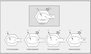
In normal conditions (Figure 1), at end expiration PL is approximately 5 cm H2O and PW is equal but opposite (­-5 cm H2O). This balance produces a Ppl of ­-5 cm H2O and an alveolar pressure (Palv) of 0 cm H2O. Therefore, there is no airflow. The contraction of the inspiratory muscles will create an expansive force of the chest wall (Pmus) which is negative in sign and which is transmitted through the pleural space to the alveoli, where the pressure becomes subatmospheric, generating a pressure gradient and thus airflow from the mouth to the alveoli. In a partial inspiration, the negative pressure generated by the inspiratory muscles exceeds PL and a subatmospheric Palv and inspiratory airflow persist. At the end of an inspiration (at total lung capacity--TLC), a balance is established between the negative pressure exercised by the inspiratory muscles and the positive pressure exerted by PW and PL. Once again Palv is 0 cm H2O and there is no airflow. Expiration begins when contraction of the inspiratory muscles decreases; thus, the elastic recoil pressures PW and PL predominate, generating a positive Palv and expiratory flow which will continue until an equilibrium of PW and PL is reached. It is now known that there is continuous but decreasing activity of the inspiratory muscles during expiration acting as a brake on expiratory flow.

Fig. 1. Schematic representation of pressure changes in different phases of the respiratory cycle in a healthy subject. PW indicates elastic recoil pressure of the chest wall; Ppl, pleural pressure; PL, elastic recoil pressure of the lung; Palv, alveolar pressure; Pmo, mouth pressure; Pmus, muscle contraction pressure. Pressures are expressed in cm H2O. The top figure (framed) represents the equivalences; the figures of partial inspiration and expiration represent the same lung volume. See explanation in text. (Adapted from Gibson.1)

In scoliosis and other chest wall disorders, the elastic properties of the lungs and airways are normal; thus, FRC or end-expiratory pressures are normal. Since there is great reduction of chest wall compliance (chest wall stiffness) in these diseases, greater inspiratory force is required to overcome the reduction without producing very great changes in Ppl. Figure 2 shows that, assuming that chest wall compliance is half of normal, PW at the same tidal volume will increase twofold over the normal level (eg, from ­-5 to +3 cm H2O, as opposed to ­-5 to -­1 cm H2O in healthy subjects) between the end of expiration and the end of inspiration. Therefore, greater inspiratory force is required to overcome chest wall stiffness.

Fig. 2. Schematic representation of pressure changes in different phases of the respiratory cycle in a subject with a chest wall disease or chest wall stiffness. See Figure 1 for equivalences. FRC indicates functional residual capacity; TLC, total lung capacity. The figures of partial inspiration and expiration represent the same lung volume. See explanation in text. (Adapted from Gibson.1)

Lung and chest wall distensibility

The lungs and the chest wall are elastic structures that tend to return to their initial shape after having been deformed by an external force. The elastic recoil pressure is that which opposes their stretch or distension. The system elasticity may be expressed as compliance and defined as volume change per unit pressure change (ΔV/ΔP).

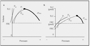
In the healthy subject a Rahn diagram (Figure 3a) illustrates that TLC is determined by the balance between the pressures generated by the respiratory muscles and the elastic recoil of the respiratory system. In a deep breath with an open glottis, Palv is zero (transrespiratory system pressure, or PRS, equals -­Pmus). In patients with chest wall diseases, which are characterized by increased chest wall stiffness (Figure 3b), PW and thus PRS will increase, meaning that balance (PRS = ­-Pmus) will occur at a much lower volume than that observed in healthy persons, such that the pressure-volume curve is flatter. Because the volume is reduced, the maximum PL is lower than normal.

Fig. 3. Pressure-volume curve determinants in a healthy subject (a) and in a subject with chest wall disease or chest wall stiffness (b). PW indicates elastic recoil pressure of the chest wall; PRS, transrespiratory system pressure; PL, elastic recoil pressure of the lung; Pmus, muscle contraction pressure; FRC, functional residual capacity; TLC, total lung capacity. Pressures are expressed as cm H2O. See explanation in text.

Evaluation respiratory muscle functions

The mechanical properties and function of the respiratory muscles can be evaluated by measuring lung volumes, chest wall motion, and pressures at different locations of the chest wall. Respiratory muscle activity can also be characterized by EMG.

In the respiratory system, force is measured as pressures, and muscle contraction is measured as volume changes or displacement of chest wall structures. To characterize the respiratory muscles quantitatively, we can use measurements of volume, displacement, pressures, and the relationships between changes in these variables over time. Thoracic geometry has an important role to play in efficiently converting force into pressure. These pressures also depend on the mechanical characteristics of the thoracic cage and the abdominal wall, with which respiratory muscles interact. A stiff chest wall resists the distortion generated by muscle action and thus permits the diaphragm to create greater pressure for a given volume change. Therefore, the pressures should be seen only as indices of the overall work of the respiratory muscles and not as a direct measure of their contraction. The global evaluation of the respiratory muscles has been dealt with in other chapters of this series; thus, we need not enter into a discussion of this particular aspect at this time.2

Kyphoscoliosis

Kyphoscoliosis is a disease of the vertebral column and its articulations, characterized by the simultaneous presence of scoliosis (lateral curvature of the spine) and kyphosis (anteroposterior spinal curvature). Signs of the latter may be destruction of vertebral bodies, bone tuberculosis, osteoporosis, etc. Primary or idiopathic kyphoscoliosis is a hereditary disease characterized by defects in the development of the vertebrae or by connective tissue disease. It accounts for 80% of cases and is more frequent in women. Secondary kyphoscoliosis generally originates in childhood neuromuscular diseases such as poliomyelitis or muscular dystrophies.3

During the course of kyphoscoliosis, the initial displacement of the spine ("primary curvature") involves two structural changes: on the one hand, a secondary curvature that develops to counterbalance the primary curve and, on the other hand, a rotation of the column on its longitudinal axis, displacing the ribs of the convex side in a posterior and outward direction to form a "hump" shape while the ribs on the concave side are constricted. The intensity of the deformity can be quantified by measuring the angles of the primary convexity. With angles greater than 100º, the deformity is considered severe and the risk of respiratory failure, alveolar hypoventilation, and cor pulmonale is very high.4

Kyphoscoliosis is expressed in functional changes with decreases in TLC, vital capacity (VC), and FRC but without changes in residual volume (RV).5,6 The severity of compression is directly related to the degree of kyphoscoliotic angulation.7-9 Diminished expiratory flows are due to the restrictive process per se, not to intrinsic airway disease.10

In advanced kyphoscoliosis, inspiratory muscle strength is significantly reduced and maximum transdiaphragmatic (Pdimax) and inspiratory (PImax) pressures are much reduced.6 In such patients, high positive gastric pressures are generated during respiration, indicating that diaphragm contraction is effective with very negative esophageal pressures. This suggests active recruitment of the intercostal muscles and the inspiratory accessory muscles. In studies of kyphoscoliosis patients, an inverse correlation has been observed between PaCO2 and PImax,, possibly indicating that inspiratory muscle restriction plays a predominant role in the development of respiratory failure in these patients.

In patients with chronic obstructive pulmonary disease (COPD) and other similar respiratory diseases, increased nerve activity has been observed in the inspiratory muscles of the rib cage and the diaphragm. Such observations suggest that a typical response to mechanical overload is involved.11 Consistent with this hypothesis, Estenne et al12 observed a PImax, a sharp decrease in dynamic lung compliance (0.04±0.03 L/cm H2O) and an increase in respiratory muscle recruitment during respiration in 8 kyphoscoliotic patients with a severe restrictive pattern (TLC 44%±8% of predicted, FRC 60%±11% of predicted, and RV 82%±20% of predicted). The observations were attributed to an automatic response of central respiratory drive.

In summary, decreased respiratory system compliance, conditioned by the stiffness which deformity transmits to the chest wall,13 is evident in adults with kyphoscoliosis. This brings about a reduction in the TLC and flattens the pressure-volume curve (Figure 3b). The respiratory load increases so that patients adopt a breathing pattern characterized by small volumes and very high respiratory rates,14 with increased dead space, alveolar hypoventilation, and disordered sleep,15 these changes being very marked at times. Similarly, the greater work of the respiratory muscles stimulates increased oxygen consumption, involving greater risk of respiratory failure due to muscle fatigue.16

Compliance in children, on the other hand, is preserved regardless of the severity of the deformity.17

Ankylosing spondylitis

Ankylosing spondylitis is an inflammatory disease of the axial skeleton that mainly affects males. Ninety-five per cent of affected individuals share the genetic marker HLA-B27. Inflammation compromises the ligamentous structures of the spinal column, the sacroiliac articulations, and the ribs, leading to the fibrosis and ossification of these structures and, thus, to an increase in rig cage stiffness due to ankylosis and fusion of the costovertebral and sternoclavicular articulations.

In spite of this rib cage stiffness, there is no very marked decrease in the lung volumes, either TLC or FRC,18 nor is there an observed correlation between the severity of the spinal angulation and the functional parameters indicating restriction. The slight reduction in lung volumes, which is possibly the result of the fusing of the ribs,19 correlates positively with disease duration and activity.20 Finally, observed decreases in both PImax and maximum expiratory pressure indicate weakness caused by intercostal muscle atrophy plus dysfunction of the diaphragm, whose activity is generally compensatory and preserved.

Pectus excavatum

Pectus excavatum is the most common deformity of the anterior thorax. Consisting of a very notable concavity of the lower third of the sternum, pectus excavatum is a congenital defect which is manifested in different degrees of severity. Thus, it is classified according to the extent of concavity visible in a lateral x-ray of the thorax. It is considered significant when the distance between the surface of the anterior wall of the thorax and the deepest part of the depression is greater than 3 cm.21 Its etiology is unknown, although a close relation to the Marfan syndrome has been found to exist, suggesting that the connective tissue around the sternum is altered. Lung volumes are at the lower limits of normal or slightly diminished, and compliance is normal unless accompanied by severe kyphoscoliosis.22

Obesity

When obesity is severe (a body mass index [BMI] greater than 30 kg/m²), lung function may be compromised. The distribution of obesity can also endanger respiratory function, independently of the total percentage of body fat.23 However, forced spirometry in patients with uncomplicated obesity is generally normal, although there may be a small decrease in forced VC, which correlates inversely with weight.24 The effects of uncomplicated obesity are generally reflected more in a significant decrease of the expiratory reserve volume, which equals FRC­RV at <=60% of predicted. This decrease has an inverse correlation with BMI. The TLC and FRC tend to be normal25 with BMI values under 45 kg/m<². Patients with obesity hypoventilation syndrome (OHS) usually manifest more severe restrictive and functional impairment, which makes it impossible to explain the adverse effects of obesity on lung function exclusively in terms of the adipose load on the chest wall.26

In patients with uncomplicated obesity, chest and respiratory system compliance is slightly diminished (approximately 80% of predicted), reaching losses of approximately 40% when OHS is present. Decreased distensibility is due to the mechanical restriction of the thorax and to increased abdominal content and fat, which place a greater elastic load and require a greater negative Ppl during inspiration.27,28 Obesity has also been related to increased airway resistance. This can be explained in part because at low lung volumes, with increased elastic tension, there is a decrease in the diameter of the small airways (a normal ratio of forced expiratory volume in 1 second to forced VC) with a consequent increase in resistance,29 a characteristic that is most pronounced in the presence of OHS.

This functional situation requires more work to overcome the elastic and resistive forces and thus leads to greater oxygen consumption by the respiratory muscles. Kress et al30 found that oxygen consumption by respiratory muscles was greater in morbidly obese patients than in normal subjects, although it decreased with mechanical ventilation. In theory, increased loading could either have a training effect or could cause fatigue if the burden of concurrent disease became very great. In uncomplicated obesity, inspiratory muscle strength is normal.

Conclusion

Diseases that constrict the chest wall lead to severe respiratory function impairment. Kyphoscoliosis, congenital disorders of the chest wall, and morbid obesity are the chest wall disorders that most commonly lead to the development of cor pulmonale and chronic respiratory insufficiency. In patients with these disorders, a stiff thorax imposes a very considerable elastic load on the respiratory muscles. This increases the work of breathing and alters lung function mechanics. If inspiratory muscle weakness is also present, the consequences may be even greater.

Even though the pathophysiology of the respiratory muscles in such disorders has not been studied to the same extent as in COPD, there is still sufficient information on functional impairment and the role it plays in the development of respiratory failure when disease progresses or when an additional burden arises from acute (eg, respiratory infection) or chronic (eg, concurrent COPD) diseases. A more detailed study of chest wall disorders will, in many cases, make it possible to modify or prevent the natural history of a disease.


Correspondence to: Dr D. Maldonado.

Fundación Neumológica Colombiana.

Calle 163.ª, 28-60. Bogotá. Colombia.

E-mail: dmaldonado@cable.net.co

Manuscript received March 24, 2003.

Accepted for publication April 2, 2003.

Bibliography
[1]
Gibson GJ..
Tests of mechanical function. In: Gibson GJ, editor. Clinical Tests of respiratory function. London: Chapman & Hall Medical.
Tests of mechanical function. In: Gibson GJ, editor. Clinical Tests of respiratory function. London: Chapman & Hall Medical, (1996), pp. 13-57
[2]
ATS/ERS statement on respiratory muscle testing..
Am J..
Respir Crit Care Med, 166 (2002), pp. 515-624
[3]
McCool FD, Rochester DF..
The lungs and chest wall diseases. In: Murray JF, Nadel JA, editors. Textbook of respiratory medicine. Philadelphia: Saunders Company.
The lungs and chest wall diseases. In: Murray JF, Nadel JA, editors. Textbook of respiratory medicine. Philadelphia: Saunders Company, (2000), pp. 2357-76
[4]
Bergofsky EH..
Respiratory failure in disorders of the thoracic cage..
Am Rev Respir Dis, 119 (1979), pp. 643-69
[5]
Weber B, Smith JP, Briscoe WA, Friedman SA, King TK..
Pulmonary function in asymptomatic adolescents with idiopathic scoliosis..
Am Rev Respir Dis, 111 (1975), pp. 389-97
[6]
Lisboa C, Moreno R, Fava M, Ferretti R, Cruz E..
Inspiratory muscle function in patients with severe kyphoscoliosis..
Am Rev Respir Dis, 132 (1985), pp. 48-52
[7]
Kearon C, Viviani GR, Killian KJ..
Factors influencing work capacity in adolescent idiopathic thoracic scoliosis..
Am Rev Respir Dis, 148 (1993), pp. 295-303
[8]
Kearon C, Viviani GR, Kirkley A, Killian KJ..
Factors determining pulmonary function in adolescent idiopathic thoracic scoliosis..
Am Rev Respir Dis, 148 (1993), pp. 288-94
[9]
Kafer ER..
Idiopathic scoliosis. Mechanical properties of the respiratory system and the ventilatory response to carbon dioxide..
J Clin Invest, 55 (1975), pp. 1153-63
[10]
Al Kattan K, Simonds A, Chung KF, Kaplan DK..
Kyphoscoliosis and bronchial torsion..
Chest, 111 (1997), pp. 1134-7
[11]
Ninane V, Rypens F, Yernault JC, De Troyer A..
Abdominal muscle use during breathing in patients with chronic airflow obstruction..
Am Rev Respir Dis, 146 (1992), pp. 16-21
[12]
Estenne M, Derom E, De Troyer A..
Neck and abdominal muscle activity in patients with severe thoracic scoliosis..
Am J Respir Crit Care Med, 158 (1998), pp. 452-7
[13]
Conti G, Rocco M, Antonelli M, Bufi M, Tarquini S, Lappa A, et al..
Respiratory system mechanics in the early phase of acute respiratory failure due to severe kyphoscoliosis..
Intensive Care Med, 23 (1997), pp. 539-44
[14]
Jones RS, Kennedy JD, Hasham F, Owen R, Taylor JF..
Mechanical inefficiency of the thoracic cage in scoliosis..
Thorax, 36 (1981), pp. 456-61
[15]
Mezon BL, West P, Israels J, Kryger M..
Sleep breathing abnormalities in kyphoscoliosis..
Am Rev Respir Dis, 122 (1980), pp. 617-21
[16]
McCool FD, Tzelepis GE, Leith DE, Hoppin FG Jr..
Oxygen cost of breathing during fatiguing inspiratory resistive loads..
J Appl Physiol, 66 (1989), pp. 2045-55
[17]
Frick SL..
Scoliosis in children with anterior chest wall deformities..
Chest Surg Clin N Am, 10 (2000), pp. 427-36
[18]
Van Noord JA, Cauberghs M, Van de Woestijne KP, Demedts M..
Total respiratory resistance and reactance in ankylosing spondylitis and kyphoscoliosis..
Eur Respir J, 4 (1991), pp. 945-51
[19]
Fisher LR, Cawley MI, Holgate ST..
Relation between chest expansion, pulmonary function, and exercise tolerance in patients with ankylosing spondylitis..
Ann Rheum Dis, 49 (1990), pp. 921-5
[20]
Feltelius N, Hedenstrom H, Hillerdal G, Hallgren R..
Pulmonary involvement in ankylosing spondylitis..
Ann Rheum Dis, 45 (1986), pp. 736-40
[21]
Kaguraoka H, Ohnuki T, Itaoka T, Kei J, Yokoyama M, Nitta S..
Degree of severity of pectus excavatum and pulmonary function in preoperative and postoperative periods..
J Thorac Cardiovasc Surg, 104 (1992), pp. 1483-8
[22]
Quigley PM, Haller JA Jr, Jelus KL, Loughlin GM, Marcus CL..
Cardiorespiratory function before and after corrective surgery in pectus excavatum..
J Pediatr, 128 (1996), pp. 638-43
[23]
Rubinstein I, Zamel N, DuBarry L, Hoffstein V..
Airflow limitation in morbidly obese, nonsmoking men..
Ann Intern Med, 112 (1990), pp. 828-32
[24]
Gibson GJ..
Obesity, respiratory function and breathlessness..
Thorax, 55 (2000), pp. 41-4
[25]
Jenkins SC, Moxham J..
The effects of mild obesity on lung function..
Respir Med, 85 (1991), pp. 309-11
[26]
Sahebjami H, Gartside PS..
Pulmonary function in obese subjects with a normal FEV1/FVC ratio..
Chest, 110 (1996), pp. 1425-9
[27]
Luce JM..
Respiratory complications of obesity..
Chest, 78 (1980), pp. 626-31
[28]
Pelosi P, Croci M, Ravagnan I, Vicardi P, Gattinoni L..
Total respiratory system, lung, and chest wall mechanics in sedated-paralyzed postoperative morbidly obese patients..
Chest, 109 (1996), pp. 144-51
[29]
Zerah F, Harf A, Perlemuter L, Lorino H, Lorino AM, Atlan G..
Effects of obesity on respiratory resistance..
Chest, 103 (1993), pp. 1470-6
[30]
Kress JP, Pohlman AS, Alverdy J, Hall JB..
The impact of morbid obesity on oxygen cost of breathing (VO[2RESP]) at rest..
Am J Respir Crit Care Med, 160 (1999), pp. 883-6
Archivos de Bronconeumología
Article options
Tools

Are you a health professional able to prescribe or dispense drugs?