


The purpose of this study was to verify the reproducibility between two different observers of an analysis method for diaphragmatic displacement measurements using direct visualization with videofluoroscopy.
Patients and methods29 mouth breathing children aged 5–12 years from both genders were analyzed. The diaphragmatic displacement evaluation was divided in three parts: videofluoroscopy with VHS recording in standing, sitting, and dorsal positions; digitalization of the images; and measurement of the distance between diaphragmatic domes during a breathing cycle using Adobe Photoshop 5.5 and Adobe Premiere PRO 6.5 software.
ResultsThe intraclass correlation coefficients presented excellent reproducibility in all positions, with coefficients always above 0.94. Mean of the measurements of the diaphagramatic domes displacement done by the two observers were similar (P<.05), with the exception of right diaphragmatic displacement in the seated position and left diaphragmatic displacement in the standing position (P<.05).
ConclusionThis study demonstrated that the evaluation of diaphragmatic displacement via videofluoroscopy is a reproducible measurement when two trained investigators are compared. This fact indicates that the present technique can be disseminated and performed by other healthcare professionals.
El objetivo de este estudio fue verificar la reproducibilidad por parte de dos observadores diferentes de un método de análisis de las mediciones del desplazamiento del diafragma con el empleo de una visualización directa con videofluoroscopia.
Pacientes y métodosSe estudió a 29 niños de ambos sexos y de entre 5 y 12 años, con respiración oral. La evaluación del desplazamiento del diafragma se dividió en tres partes: videofluoroscopia con grabación mediante VHS en las posiciones de bipedestación, sedestación y decúbito supino; digitalización de las imágenes; y medición de la distancia entre las cúpulas diafragmáticas durante un ciclo de respiración, con el empleo de los programas informáticos Adobe Photoshop 5.5 y Adobe Premiere PRO 6.5.
ResultadosLos coeficientes de correlación intraclase mostraron una excelente reproducibilidad en todas las posturas, con valores de los coeficientes situados siempre por encima de 0,94. La media de las mediciones del desplazamiento de las cúpulas diafragmáticas realizadas por dos observadores fue similar (P<0,05), con la excepción del desplazamiento diafragmático derecho en posición de sedestación y el desplazamiento diafragmático izquierdo en posición de bipedestación (P<0,05).
ConclusiónEste estudio puso de manifiesto que la evaluación del desplazamiento diafragmático mediante videofluoroscopia es una medición reproducible cuando se comparan los resultados obtenidos por dos investigadores adecuadamente preparados para ello. Este hecho indica que la técnica evaluada puede ser difundida y pueden aplicarla otros profesionales de la asistencia sanitaria.
The diaphragm is the main respiratory muscle and it contracts during inspiration, generating a caudal movement that is proportional to the intrathoracic pressure and tidal volume.1 The analysis of the diaphragmatic excursion provides a method for evaluating the degree of muscle deterioration in neuromuscular and respiratory diseases. In recent years, different radiological methods have been used to evaluate the diaphragmatic excursion, among these ultrasound, sonomicrometry, electromyography, nuclear magnetic resonance and videofluoroscopy.2–6 Ultrasound provides a subjective evaluation of the muscle movement.2,7 Sonomicrometry measures the distance between pairs of piezoelectric crystals with the use of acoustic signals.8 Electromyography monitors the electrical activity in the muscle.3 Nuclear magnetic resonance provides satisfactory effectiveness in the determination of the morphology and the motility of the diaphragm.5
Videofluoroscopy occupies an important place among the methods for measuring the distance of the diaphragmatic excursion during breathing as it allows for the visualization of the entire hemi-cupula and some devices simultaneously examine the right and left cupulas. Videofluoroscopy allows for qualitative evaluation of muscle motility, as well as quantitative evaluations to measure the excursion or the size of the diaphragmatic cupulas.5,9 This method can be used to evaluate the paralysis caused by phrenic nerve injury,10 the presence of malformations,6 muscle contraction after electrical stimulation,5 the movement of the esophagus, which is produced in synchronicity with the diaphragm,2 the weakness of the diaphragm in patients with sleep disorders,4 motility after thoracotomy or heart surgery7 and the correlation between the tidal flow and the contraction of the diaphragm.11
We have recently reported the results obtained in the fluoroscopic evaluation of the diaphragm movement in children with oral breathing. The method was relatively simple and easy to use. As this system uses video recording, the images can be examined as frequently as necessary without the need to expose the patient to more X-ray radiation.6 However, as occurs with the majority of the diagnostic tests, there may be discrepancies between the results obtained by different evaluators. In order for a diagnostic test to be considered adequate, it should produce similar results (reproducibility) when applied by different evaluators. Consequently, the determination of the existing coincidence between the results obtained by two different observers is fundamental for determining the reproducibility of an exploration.
The objective of the present study was to evaluate the reproducibility of fluoroscopy by means of the direct visualization of the images recorded by video for the determination of the excursion of the diaphragm in children with mouth breathing using the level of coincidence between two blinded evaluators.
Patients and MethodsWe evaluated a non-probabilistic, opportunity sample made up of 29 children who were being treated in the Mouth Breathing Outpatient Clinic of the Otorhinolaryngology Division at the Universidade Federal de São Paulo (Brazil). All the procedures of the study were previously approved by the Institutional Review Council at the university (protocol 1611/03) and the parents of the children gave their written informed consent. The inclusion criteria were the following: children with oral breathing, boys and girls aged 5-12. The sample size was estimated based on reproducibility studies found in the literature.12,13
Evaluation of Diaphragmatic ExcursionThe following steps were followed to evaluate diaphragmatic excursion: (1) fluoroscopy with video recording of the breathing cycles; (2) examination of the images; and (3) measurement of the excursion of the diaphragm cupula during calm breathing.
FluoroscopyBefore the test, the patients remained sitting quietly for at least 15min. Videofluoroscopy was performed with an X-ray apparatus (Medicor model 750B), an image intensifier and a standard video screen (2:1 with 525 lines and 60Hz) from which the images were transferred to a videotape (Philco model 7400). We set a metallic object measuring 0.1cm by 2.0cm at the level of the breast nipples, bilaterally, with the aim of allowing their visualization in the video recording. This strategy made it possible to apply a comparative and proportional equation between the excursion distance and the length of the metal piece.
The images of the diaphragmatic excursion on the right side were registered first, followed by the left side. We recorded four cycles of calm breathing for each cupula while standing, sitting and in supine decubitus position, with an X-ray apparatus and anteroposterior (AP) projection. The feet were parallel in all the positions of the exam, and the arms were placed next to the body in the standing and supine decubitus positions. In the sitting position, the participants were indicated to rest their hands on the middle third of the inside of each leg. The children were indicated to keep their eyes open, looking at the horizon. There was no verbal interference for posture correction. The torso remained uncovered and we avoided compression of the participants trousers in the abdominal area.14 Out of the four cycles registered bilaterally in each position, we selected for analysis the one that had a more harmonious movement. We dismissed the cycles in which there were sudden torso movements, cough, sneezing, reflex movements or speech. The excursion of the diaphragm registered on video was measured independently by two observers who did not know the measurements made by the other evaluator, applying the method that is described further ahead.
Digitalization of the Images and Measurement of the Distance Between the Diaphragm CupulaThe following equipment was used for the analysis of the diaphragmatic excursion distance: (1) a microcomputer with an image capturing card (Pinnacle model PRO ONE RTDV); (2) an SVHS video recorder (Sony model 5800); (3) a video screen (Sony TRINITON model 1351); (4) a video controller (Sony model PVE 500). The analyses were done in the following order, using the corresponding software.14
- a)
Two still images: one with the highest point and another with the lowest point reached by the diaphragmatic cupula during a respiratory cycle (Fig. 1) (Adobe Premiere PRO 6.5).
 arrow: metallic object; (B) white L-shaped lines: vertebra position; (C) lower horizontal line: cupula of the diaphragm during inspiration; (D) upper horizontal line: cupula of the diaphragm during expiration; (E) small vertical line between the two horizontal lines: excursion of the diaphragm cupulas during calm breathing (see the text for more information).") Fig. 1.
Fig. 1.Measurement of the excursion of the diaphragmatic cupula during calm breathing. (A) arrow: metallic object; (B) white L-shaped lines: vertebra position; (C) lower horizontal line: cupula of the diaphragm during inspiration; (D) upper horizontal line: cupula of the diaphragm during expiration; (E) small vertical line between the two horizontal lines: excursion of the diaphragm cupulas during calm breathing (see the text for more information).
- b)
For the simultaneous visualization of two superimposed images, the transparency of the image with the highest point reached by the diaphragm cupula was set at 40%, and corresponded with the upper limit of excursion (Fig. 1) (Adobe Photoshop 5.5).
- c)
The images were superimposed using the right angle created by a specific vertebra that was used as a reference point for both images.
- d)
The highest and lowest points reached by the diaphragm cupulas were identified by means of horizontal lines (Fig. 1) (Adobe Photoshop 5.5).
- e)
The excursion distance was measured (Fig. 1) (Adobe Photoshop 5.5).
- f)
The longitude of the metal object set at the level of the nipple was measured (Fig. 1) (Adobe Photoshop 5.5).
- g)
The real excursion distance was calculated (x) with the use of the following formula:
 arrow: metallic object; (B) white L-shaped lines: vertebra position; (C) lower horizontal line: cupula of the diaphragm during inspiration; (D) upper horizontal line: cupula of the diaphragm during expiration; (E) small vertical line between the two horizontal lines: excursion of the diaphragm cupulas during calm breathing (see the text for more information).")
x=distance measured between the diaphragm cupulas (E) × 2cm/length of the metal object visualized in the image (A).
Statistical AnalysisThe numerical data are expressed as means and standard deviation. The data of the categorical variables are expressed in absolute values and percentages. The two observers evaluated the excursion of the diaphragm independently and blind to the results of the other. Later, the data were compared. The Student's t-test was used for paired data in order to compare the means and differences between the measurements made by the two examiners. The interclass correlation coefficient and the 95% confidence interval were used to evaluate the reproducibility of the measurements. A level of statistical significance of 5% was adopted.
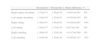
ResultsTable 1 shows the demographic data of the participating children. The mean values for age, weight and height were 8 years, 29.7kg and 1.30m, respectively.
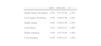
Table 2 presents the mean of the diaphragm excursion values calculated by the two evaluators, as well as the mean differences between them. The excursion of the right diaphragm in the sitting position and the excursion of the left diaphragm in the standing position were the only measurements where discrepancies were identified between the two evaluators (P<.05). The mean differences between the two measurements were 2.6 and 6.6mm, respectively (Table 3). However, the intraclass correlation coefficient showed an excellent reproducibility in all the positions, with coefficients that were always above 0.94 (Table 3).
Excursion of the diaphragm cupulas during calm breathing, in centimeters.
| Researcher 1 | Researcher 2 | Mean difference | P | |
| Right supine decubitus | 1.35±0.53 | 1.36±0.50 | −0.007±0.081 | NS |
| Left supine decubitus | 1.34±0.59 | 1.35±0.61 | −0.010±0.055 | NS |
| Right sitting | 1.46±0.61 | 1.48±0.63 | −0.026±0.067 | .044 |
| Left sitting | 1.39±0.57 | 1.37±0.50 | 0.027±0.180 | NS |
| Right standing | 1.30±0.45 | 1.32±0.46 | −0.017±0.069 | NS |
| Left standing | 1.24±0.46 | 1.30±0.46 | −0.065±0.147 | .023 |
Mean±standard deviation; NS: not significant; P: level of significance.
Intraclass correlation coefficient values of the readings of the excursion del diaphragm during calm breathing between two different observers.
| ICC | 95% CI | P | |
| Right supine decubitus | 0.98 | 0.97-0.99 | <.001 |
| Left supine decubitus | 0.99 | 0.99-0.99 | <.001 |
| Right sitting | 0.99 | 0.98-0.99 | <.001 |
| Left sitting | 0.94 | 0.88-0.97 | <.001 |
| Right standing | 0.98 | 0.97-0.99 | <.001 |
| Left standing | 0.94 | 0.86-0.97 | <.001 |
ICC: intraclass correlation coefficient; CI: confidence interval; P: level of significance.
In recent years, different radiological methods have been used to evaluate diaphragmatic excursion, among them ultrasound, sonomicrometry, electromyography, nuclear magnetic resonance and videofluoroscopy.2–6 Ultrasound is a good method for the subjective evaluation of the muscle movement, but it does not allow for the visualization of a complete hemi-cupula.2,7 Sonomicrometry measures the distance between pairs of piezoelectric crystals with the use of acoustic signals. In recent articles, the use of this method has been reported for determining the size of heart chambers and measuring the contraction time of the diaphragm. All the experiments in which this method has been used to date have been carried out in animal models.8,11,15 Electromyography monitors the electrical activity in the muscle. The surface electrodes placed on the chest evaluate simultaneously other respiratory muscles. Needle electrodes are necessary for the isolated evaluation of the diaphragm.4 In some studies catheters have been introduced in the esophagus for evaluation.7 Nuclear magnetic resonance offers satisfactory effectiveness in the evaluation of the diaphragm, but it is expensive and could be replaced by a method with lower cost.5
Videofluoroscopy occupies an important place among the methods for measuring diaphragm excursion distance during breathing, as it enable the visualization of the entire hemi-cupula and some devices allow for the right and left cupulas to be examined simultaneously. Moreover, according to Braun et al. (1995), the exposure to X-ray radiation for 5min causes no damage to patients. Videofluoroscopy enables us to make qualitative evaluations of the muscle motility, as well as quantitative evaluations for measuring the excursion or the size of the diaphragm cupulas.5,9 This method can be used to evaluate the paralysis caused by a lesion of the phrenic nerve,15 presence of malformations,6 muscle contraction after electrical stimulation,5 movement of the esophagus produced synchronically with the diaphragm,2 weakness of the diaphragm in patients with sleep disorders,4 motility after thoracotomy or heart surgery7 and the correlation between the tidal volume and the contraction of the diaphragm.11
The present study reveals that the measurement of diaphragmatic excursion with the use of direct visualization with fluoroscopy and a later analysis of the video recording is a reproducible technique when the recording is analyzed by two different evaluators. These results indicate that the method can be taught, is simple to use and does not require much time. It had been used previously, but with no confirmation of its reproducibility.14 As the tests are recorded on video, which allows for later analysis, the patients are exposed to less X-ray radiation,16 and the recording can also be made in a digital format on digital versatile disc (DVD).
The reliability or reproducibility is defined as the level of coincidence between two measurements. The reliability can be evaluated when a measurement is done by two different observers or when two measurements are done by the same evaluator at different moments. It is an important step in the process of developing a method, as it measures the capacity of obtaining the same result in two different readings. The reliability of the continuous variables should be evaluated with the use of the intraclass correlation coefficient (ICC), which reflects the level of coincidence between two evaluations of a same observer on two different occasions.17 As occurs with many other physiological determinations in humans, there could be a certain variation between the two determinations, even though the patient is considered stable. In the reliability studies, a smaller degree of variation between the values obtained in two different evaluations leads to a higher ICC, which indicates a greater reliability of the determination. A measurement is considered reliable when the ICC is higher than 0.70 (1.00 indicates perfect reproducibility).17 As shown in Table 3, the ICC of all the determinations was always above 0.94. Thus, this present study manifests that it is possible to convert an apparent diaphragm excursion measurement into a real distance by means of a simple equation that takes into account the proportion of the measurement of the diaphragmatic excursion and the length of a metallic object placed at the level of the nipples.
At rest, the diaphragm moves around 1cm, while during forced inspiration and expiration this movement can reach 10cm. Variations from the normal pattern may indicate a neuromuscular disease.18 Videofluoroscopy could be used in longitudinal studies, as long as there is an indication for the study in the patient, as in the case of the individuals with neuromuscular diseases.
The participants in the present study were exposed to a single dose of radiation for 1min. According to Lederman et al. (2002), this length of exposure with the X Medicor X-ray apparatus (model 750B) causes no damage in patients. In a study of patients who underwent diaphragm videofluoroscopy with a 5min exposure to radiation, Braun et al. (1995) indicated that, when the quantity of radiation calculated over the exposure time is less than 12.5Rads (a unit of radiation dose absorbed, largely obsolete), the patient suffers no injury.
Braun et al. (1995) carried out a qualitative evaluation of diaphragmatic excursion by simultaneously using videofluoroscopy, spirometry and plethysmography. It would be useful to know the correlation between the tidal volume and the excursion of the diaphragm measured with videofluoroscopy. However, as this was not the objective of the present study, the tidal volume was not determined. We recommend further studies in this direction.
Kolar et al. (2007) analyzed diaphragmatic excursion with the use of a computer program designed exclusively to this end which examines the diaphragm in a sagittal projection. Nevertheless, this program has restricted access. The program that we have used in this study (Adobe Photoshop 5.5) is widely accessible and can be used by those who have standard knowledge of its use.
Regarding the interobserver coincidence, there were no statistically significant differences in the readings (Table 2), except for the measurements of right diaphragmatic excursion in the sitting position and left diaphragmatic excursion while standing. Given the fact that these differences were small and observed in only two positions, videofluoroscopy can be considered very reliable.
ConclusionThe present study shows that the method proposed for evaluating diaphragmatic excursion by fluoroscopy is reproducible. Furthermore, the method can be easily taught and assimilated and does not require much time. There are numerous situations in which this method can be used, such as neuromuscular diseases, respiratory diseases and cases in which an apparent cause for dyspnea cannot be identified.
Conflict of interestThe authors declare having no conflict of interests.
Please cite this article as: Yi LC, et al. Fiabilidad de un método de análisis para medir el desplazamiento del diafragma mediante visualización directa con videofluoroscopia. Arch Bronconeumol. 2011;47:310-4.


