


Sjögren's syndrome (SS) is a chronic inflammatory autoimmune disease characterized by lymphocytic infiltration of the exocrine glands, particularly the lacrimal and salivary glands, causing the characteristic symptoms of xerophthalmia and xerostomia. The lung, thyroid, kidney and the hepatobiliary tract can also be affected. This disease can be present in an isolated form (primary SS), or it can be associated with other connective tissue diseases (secondary SS). Pleural effusion (PE) in primary SS is rarely described in the literature.1
We report the case of a 40-year-old woman with insulin-dependent diabetes mellitus, consulting due to xerostomia and xerophthalmia for some months, and intermittent diffuse arthromyalgia. Vital signs were normal and the only notable finding on physical examination was limited mobility of the right shoulder. Routine clinical laboratory tests (including angiotensin-converting enzyme levels) and urine tests were normal. Of particular interest were rheumatoid factor 103IU/ml (upper level of normal 14IU/ml), positive antinuclear antibodies (ANA) (1/640), and positive anti-Ro/SS-A and anti-La/SS-B antibodies and immunoglobulin G 4050mg/dl. The Schirmer test was positive in both eyes, a minor salivary gland biopsy showed Chisholm grade 2 lymphocytic infiltration, and scintigraphy of the salivary glands was consistent with SS. A diagnosis of primary SS was established on the basis of these results.
Four years later, the patient developed right pleuritic pain, without dyspnea, cough or expectoration, but with low-grade fever in the evenings. She reported no previous chest injury and no contact with tuberculosis patients. Physical examination was unremarkable, except for signs of right PE. The chest computed tomography revealed thick-walled cystic and cavitary lesions in the right lung and a small ipsilateral PE. Fiberoptic bronchoscopy was performed that was macroscopically normal, and smear tests and PCR for Mycobacterium tuberculosis in bronchoalveolar lavage were negative. PE was an exudate (total pleural fluid protein/serum protein ratio 0.7; lactate dehydrogenase 597IU/l and pleural fluid LDH/serum LDH 1.8), leukocytes 5830/μl (neutrophils 14%, lymphocytes 24%, eosinophils 16%, macrophages 46%), with normal pH, glucose, amylase, adenosine deaminase, tumor markers and N-terminal pro-brain natriuretic peptide levels, rheumatoid factor 58.6IU/ml, positive ANA (1/320), and positive anti-Ro/SS-A and anti-La/SS-B antibodies. Bacteriological cultures, cultures for Mycobacterium tuberculosis, and cytology for malignancy were negative. The tuberculin skin test was positive (12mm). Lung biopsy (follicular bronchiolitis with areas of lymphoid interstitial pneumonia and numerous plasma cells) and pleural biopsy (infiltration by mononuclear cells) were performed using video-assisted thoracoscopy. PE resolved after surgery, and did not reappear during the 2 years of follow-up.
The diagnosis of SS is established on the basis of 6 criteria2: 2 subjective (eye and mouth symptoms), and 4 objective criteria that include ocular and oral symptoms and histopathological and serological findings (ANA and anti-Ro/SS-A or anti-La/SS-B antibodies). At least 4 of the 6 criteria (including histopathological and serological findings) or 3 of the 4 objective criteria are required for a diagnosis of SS. Our patient presented with ocular (xerophthalmia) and oral problems (xerostomia), positive Schirmer test, lymphocytic infiltration of the minor salivary gland (with consistent salivary scintigraphy), and positive anti-Ro/SS-A and anti-La/SS-B antibodies. No evidence was found of other connective tissue diseases, such as rheumatoid arthritis, systemic lupus erythematosus, or scleroderma, confirming the diagnosis of primary SS.
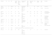
PE is very unusual in primary SS. A review of 4 series including 487 patients found that only 5 SS subjects had PE (1%), and of these 3 were secondary SS (2 had rheumatoid arthritis and 1 had systemic lupus erythematosus).3,4 However, in a study of 573 patients with primary SS, 5.7% (30/522) had PE, although the authors did not provide any more details.5 Only 10 cases of primary SS and PE have been reported in the English-language literature,6–15 although 1 case appears to be caused by concomitant heart failure.15 Although no criteria are available to define when PE is due to primary SS, they generally all show common characteristics: uni or bilateral PE corresponding to lymphocytic exudates, with a nucleated cell count of 1500–11,000/μl and normal pH, glucose and adenosine deaminase (Table 1). Anti-Ro/SS-A antibodies, rheumatoid factor and ANA were not always performed to establish the diagnosis (6/9 [66.7%]; 4/9 [44.4%, negative in 2] and 3/9 [33.3%], respectively). Pleural biopsy is nonspecific and only shows lymphocytic infiltrate. PE progress is generally favorable, since most patients respond to corticosteroids, and it may even resolve spontaneously. In our case, the pleural fluid results confirmed that PE was due to SS. PE was predominantly eosinophilic (16%), a finding not previously described, although the mononuclear cell count was also high (70%). The predominance of eosinophils does not seem to be attributable to air or blood entering the pleural space, since the procedure was not traumatic, the fluid obtained was serous, and only a thoracocentesis was performed. Analytical data from the fluid and the duration of follow-up rule out the possibility that PE originated from another source (cancer, parapneumonic, or tuberculosis, the most frequent causes of pleural exudate in our setting). The patient did not develop any other connective tissue disorder during the period that could have been responsible for the PE. PE resolved spontaneously, with no need for corticosteroids, as described previously.6
Characteristics of Reported Cases of Primary Sjögren's Syndrome With Pleural Effusion.
| Reference | Sex | Age | Side | Exudate (Light criteria) | Total Nucleated Cell Count (cells/μl) | Nucleated Cell Count (%) | Anti-SSA and Anti-SSB Ab | Rheumatoid Factor (IU/ml) | ANA | Glucose (mg/dl) | ADA (U/l) | Resolution |
|---|---|---|---|---|---|---|---|---|---|---|---|---|
| Alvarez-Sala et al.6 | F | 64 | Bilateral | Yes | ND | Predominantly lymphocytes | ND | – | ND | Normal | ND | Spontaneous |
| Ogihara et al.7 | M | 62 | Right | Yes | 2600 | 39% lymphocytes, 57% monocytes | + | ND | ND | 131 | Normal | Corticosteroids |
| Suzuki et al.8 | F | 53 | Left | Yes | ND | ND | ND | ND | ND | ND | ND | Corticosteroids Cyclophosphamide |
| Kawamata et al.9 | M | 70 | Left | Yes | 11,000 | 99% mononuclear cells | + | 80.9 | ND | ND | ND | Corticosteroids |
| Horita et al.10 | M | 73 | Bilateral | Yes | 2400 | More lymphocytes that neutrophils | + | ND | ND | 88 | ND | No response to corticosteroids Intrapleural tetracycline |
| Teshigawara et al.11 | M | 65 | Bilateral | Yes | 1520 | 84% Lymphocytes | + | – | + | ND | ND | Corticosteroids |
| Makimoto et al.12 | M | 63 | Bilateral | Yes | ND | Predominantly lymphocytes | + | ND | + | ND | ND | Corticosteroids |
| Ohe et al.13 | F | 58 | Bilateral | Yes | ND | ND | ND | ND | ND | 89 | Normal | Corticosteroids Albumin Diuretics |
| Ma et al.14 | F | 42 | Bilateral | Yes | 5000 | 98% mononuclear cells | + | 75.3 | + | ND | 12 | Hydroxychloroquine |
| Yamasaki et al.15 | F | 49 | Right | Associated congestive heart failure. No pleural fluid biochemical data | ||||||||
| This study | F | 44 | Right | Yes | 5830 | 16% eosinophils 24% lymphocytes 46% macrophages | + | 58.6 | + | 111 | 44 | Spontaneous |
Ab: antibodies; ADA: adenosine deaminase; ANA: antinuclear antibodies; F: female; M: male; ND: not determined; (+): positive; (–): negative.
In conclusion, despite the fact that the PE is a rare manifestation of primary SS, it should be considered in patients with this disease in which the etiology of the PE is not clear. Nevertheless, a definitive diagnosis requires not only certain biochemical characteristics, but also a long follow-up period to rule out other causes.
Please cite this article as: Ferreiro L, San José E, Suárez-Antelo J, Valdés L. Síndrome de Sjögren primario con derrame pleural. Arch Bronconeumol. 2017;53:598–600.


