


Dermatofibrosarcoma protuberans (DFSP) is a rare tumor of the soft tissue that is locally aggressive. Metastases are extremely uncommon and there are few cases published in the literature.1–4 We report the exceptional case of a mediastinal mass as the unique presentation of metastatic disease of a DFSP diagnosed by blind transbronchial needle aspiration (TBNA) with a histology needle.
The patient is a 50-year-old male, a smoker, who in 2004 had been diagnosed with DFSP that was 7cm in diameter and located in the left scapulohumeral region. The initial treatment was surgical, but after the resection the deep edges were affected. The patient had several local relapses that were treated surgically in 2006 and 2009. In the last surgical biopsy, the edges were tumor-free and the patient received no adjuvant treatment. Recently, the patient came for a consultation due to fever, arthralgia and unproductive cough. Radiography showed a paratracheal mass. CT confirmed the presence of a heterogeneous mediastinal mass with compression of the adjacent structures (Fig. 1). Bronchoscopy revealed edematous mucosa at the entrance of the main bronchus and the upper right lobe. Blind TBNA was performed with an MW-319 histology needle (Bard-Wang, Billerica, MA, USA) and the anatomopathologic result was DFSP metastasis, based on the morphology of the biopsies and supported by immunohistochemistry techniques (strong expression of vimentin and CD34) and by the genetic-molecular study (presence of translocation of chromosomes 17 and 22).
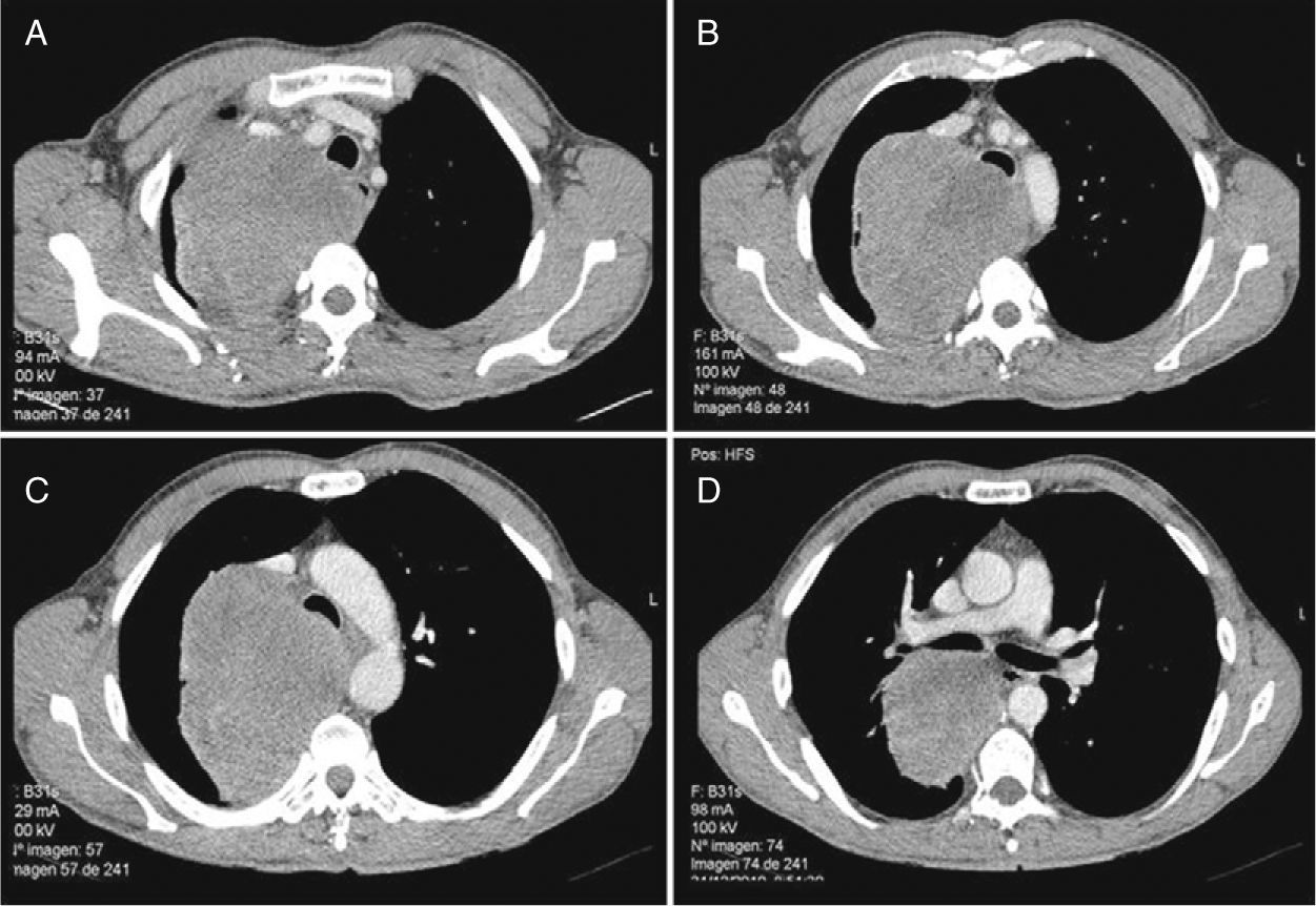
In the computed tomography images, one can observe in the middle mediastinum a large mass measuring 12×9.7cm that is heterogeneous, with well-defined edges and hypodense areas in its interior (A). The lesion compresses the trachea and displaces it towards the left (B and C) and it extends up to the entrance of the right upper bronchus, displacing it and compressing the end of the bronchus and the parenchyma of the upper right lobe (D).
DFSP is characterized by its limited metastatic potential, but it has a high risk for local recurrence.2,3 Approximately 10% of DFSP can transform into fibrosarcoma, which is a more aggressive form with greater metastatic potential.2,3 The appearance of distance metastasis is rare.1–4 In the literature, there are very few cases reported.2,3 Rutgers et al.3 only found 1% of metastasis in regional lymph nodes and 4% distance metastasis, fundamentally in the lungs.3 In one of the more extensive studies,2 only 5 out of 218 patients presented distance disease, but in no case was there affectation of the mediastinum. In two of these patients, there were no risk factors for metastatic disease, such as the affectation of the surgical margins after tumor excision or the existence of a sarcomatous component.2 In our case, the patient had presented two local recurrences, but no fibrosarcomatous component in the histological studies. The presentation of metastatic DFSP as a single mediastinal mass is exceptional, which, in addition to the method of diagnostic confirmation used, makes it especially worthy of reporting. Blind TBNA can obtain samples of mediastinal or hilar lesions and the larger-caliber needles obtain cylinders of tissue in cases of suspicion for lymphoma, other primary or metastatic non-epithelial neoplasms and granulomatous diseases.5,6 In our patient, histological TBNA enabled us to obtain sufficient tissue sample to be able to establish the morphological diagnosis of metastatic DFSP and carry out proper molecular determinations and immunohistochemistry techniques.
In conclusion, DFSP is a rare malignant dermal neoplasm characterized by slow infiltrative growth and low potential for metastasizing, but with a tendency towards recurring locally. In repeatedly recurring tumors, the risk for transformation into a more malignant form is higher. Therefore, although metastases are extremely rare, the presence of a mediastinal mass should be included in the differential diagnosis of patients with a history of DFSP, while conventional TBNA with a histological needle could be a good diagnostic option.
Please cite this article as: Lourido Cebreiro T, et al. Dermatofibrosarcoma metastásico, una causa poco frecuente de masa mediastínica. Arch Bronconeumol. 2011;47:577–8.


