
We have read with great interest the recently published article by Dr. Singh and Dr. Bal1 about lung cancer in heavy smokers that presents as symptomatic solitary lung cysts. Their article shed new light on lung cysts as a common radiological manifestation of squamous carcinoma of the lung on computed tomography (CT).
Earlier studies had suggested that lung cyst lesions could be an initial radiological sign of bronchogenic carcinoma on thoracic CT. According to Lan et al.,2 their patient was a 27-year-old woman who did not smoke, while the Singh and Bal patient was a 45-year-old male who was a heavy smoker. Both cases describe lung cancer that presents as solitary cystic lesions in the lower lung lobes. In contrast, the histologic pattern of the lung cancer was an adenocarcinoma in the case of Lan et al., while it was only a squamous carcinoma in the present case. More recently, we have identified disseminated thin-walled cystic lesions as a new radiographic sign of pulmonary adenocarcinoma.3 In our case, we have reported a 39-year-old male who was a non-smoker with the growth of a centrally located mass in the upper left lobe and rapidly progressive diffuse cystic lesions in both lungs. We also observed hilar and mediastinal lymphadenopathies as well as an elevation in the tumor markers in circulating blood (CEA and CA-125). We performed a transbronchoscopic lung biopsy together with histologic tests and immunohistochemistry. These data have finally established the diagnosis of pulmonary adenocarcinoma.
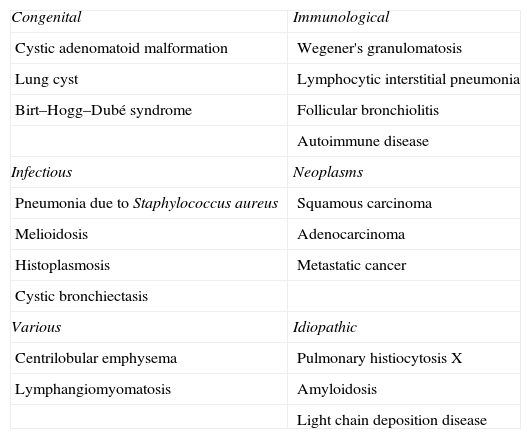
What make these cases interesting are not only their uncommon forms of presentation but also their diagnostic difficulties. Cystic lung lesions, they being either solitary or diffuse, are mainly observed in benign disorders. However, other authors and we have demonstrated the presence of cystic lung lesions as being signs of malignant thoracic diseases.1–5 The diagnostic challenge arises from the prevalence of cystic lung lesions in several thoracic disorders (Table 1). Thus, although it is not frequent, the presence of cystic lung lesions can also be associated with malignant diseases. The access to lung biopsy can be key in the precise diagnosis of these patients.
Causes of Cystic Lung Lesions.
| Congenital | Immunological |
| Cystic adenomatoid malformation | Wegener's granulomatosis |
| Lung cyst | Lymphocytic interstitial pneumonia |
| Birt–Hogg–Dubé syndrome | Follicular bronchiolitis |
| Autoimmune disease | |
| Infectious | Neoplasms |
| Pneumonia due to Staphylococcus aureus | Squamous carcinoma |
| Melioidosis | Adenocarcinoma |
| Histoplasmosis | Metastatic cancer |
| Cystic bronchiectasis | |
| Various | Idiopathic |
| Centrilobular emphysema | Pulmonary histiocytosis X |
| Lymphangiomyomatosis | Amyloidosis |
| Light chain deposition disease |
The present study was funded by grants from the National Natural Science Foundation of China (Number 81073107).
Please cite this article as: Ye M-X, et al. Quistes pulmonares como manifestación radiológica de enfermedades benignas y malignas: errores en el diagnóstico. Arch Bronconeumol. 2012; 48: 138.


