


A 74-year-old man presented in the emergency room in November 2012 with bloody expectoration, having expelled a solid “foreign body” of a fleshy appearance. He reported that in the days leading up to the event, he had experienced a sensation of self-limiting wheezing without dyspnea. The patient had no history of smoking or substance or alcohol abuse. In 2003, he required hospital admission for pneumonia and, in April 2008, underwent right upper lobectomy for a typical bronchial carcinoid tumor. At the time of presentation, he was attending regular check-ups and remained free of recurrence.
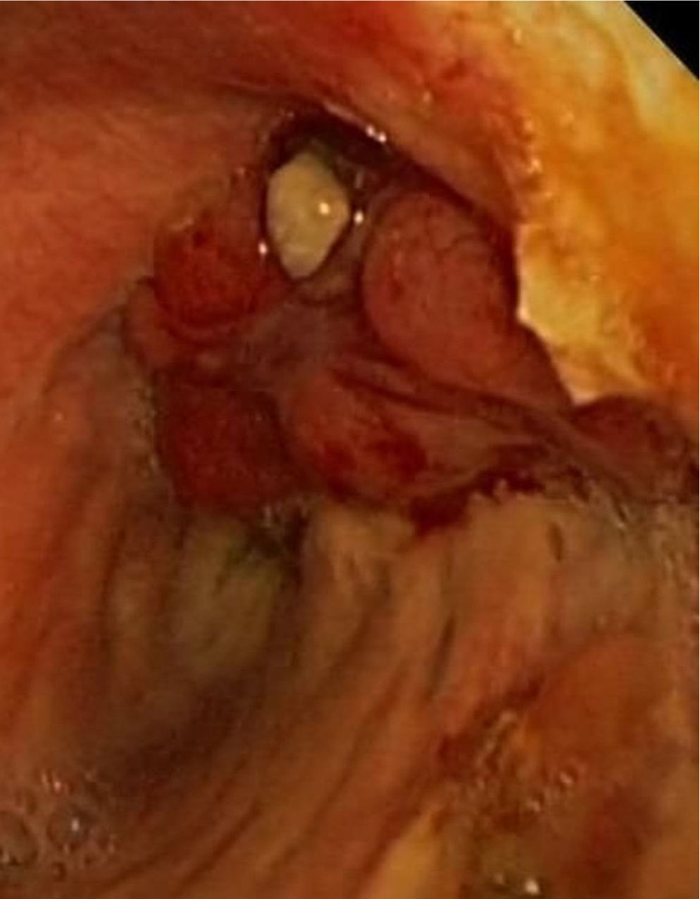
Physical examination revealed wheezing in the right hemithorax, with no other findings. Clinical laboratory tests showed hemoglobin 11.1g/dl; all other parameters were normal. The chest X-ray revealed a loss of right hemithorax volume due to the right upper lobectomy, but was unchanged compared to previous images. Computed tomography (CT) of the chest and abdomen showed polypoid lesions in the right main bronchus and in the stump of the right upper lobar bronchus. On bronchoscopy, bloody remnants and several rounded, hypervascular, millimeter-sized lesions were observed in lower third of the trachea and right anterolateral and anterior wall of the right main bronchus, above the entrance to the middle lobe. The carina and left bronchial tree were normal. A lobulated mass was observed in the area of the right upper lobectomy stump, suggestive of carcinoid tumor (Fig. 1). The bronchial biopsy results reported a well-differentiated carcinoid type lesion with an organoid growth pattern and no necrotic foci that was determined to be a high grade carcinoma due to the 70% proliferation index. Immunohistochemistry showed positive Ki-67 expression in 70% of the intensely synaptophysin-positive tumor cells. The extension studies were completed with an octreotide scintigraphy that did not show any pathological radiotracer uptake.1
In the Chest Surgery Department, the tumor tissue was locally excised with argon gas cryoablation of the lesions, with the exception of the most distal infiltration of the right and intermediate bronchus. The patient then received systemic chemotherapy.2
Carcinoid tumor is a malignant neuroendocrine carcinoma originating in the glandular basal enterochromaffin cells (“Kulchitsky cells”). The most common locations are gastrointestinal (55%) and pulmonary (30%). In the lung, it occurs most frequently in endobronchial regions of the main or lobar bronchi (70%). Histological subtyping categorizes these tumors as typical carcinoid (very slow growth and 4 times more common) and atypical carcinoid, depending on the number of mitosis per field and the presence of necrosis.3
The clinical presentation is often asymptomatic or it can present with bleeding and obstruction, causing cough, wheezing, chest pain or recurrent pneumonia. In only 5% of cases is there secretion of the vasoactive substances responsible for the carcinoid syndrome. It is diagnosed by chest X-ray, CT and bronchoscopy. Magnetic resonance imaging and somatostatin receptor scintigraphy are used for staging.4
Treatment modalities include elective radical surgical, endobronchial excision by laser, somatostatin analogs, interferon alfa, chemotherapy and radiotherapy. Five-year survival rates for the typical subtype is 82%–87% and that for the atypical subtype is 56%–75%.5
This case is of interest because, despite the advanced age of onset (69 years) and a diagnosis of typical carcinoid tumor, there was local recurrence after the patient was disease-free for more than 4 years. The tumor showed no necrotic element but the mitotic index was high, so the final diagnosis was atypical carcinoid carcinoma. The central location of the recurrence prevented radical treatment. The patient is currently asymptomatic, leading a normal life and attending regular check-ups.
Please cite this article as: Santalla Valle EA, García S, Cartón MB. Recidiva local de tumor carcinoide bronquial. Arch Bronconeumol. 2014;50:81–82.


