


Ascariasis is the most common helminthic infection. However, helminthic involvement of the pleura is very rare. We report a case of an ascaris worm found in needle-guided pleural percutaneous aspiration.
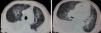
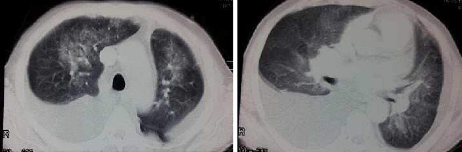
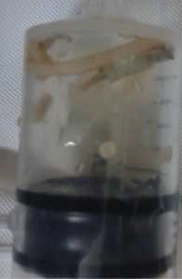
A 60 year-old male, smoker, presented with a 1-month history of dyspnea of gradual onset and progressive course, and hemoptysis. The condition was associated with anorexia, weight loss, and abdominal discomfort. Physical examination showed splenomegaly and normal temperature, with nolymphadenopathy or hepatomegaly. Respiratory examination showed dullness on percussion over the right and left lung base with decreased breath sounds. The rest of the examination was unremarkable. A chest radiograph showed bilateral pleural effusion and bilateral pulmonary infiltrates in both lung fields. A computed tomographic (CT) scan showed bilateral pleural effusion and bilateral consolidation involving upper lobes, middle lobe, and lingula (Fig. 1). White cell count 96000 with 75% eosinophils. Abdominal ultrasound showed splenomegaly with simple splenic cyst. Stool examination revealed amebiasis. A repeat stool examination showed ascaris ova. Based on the clinical and laboratory findings, diagnosis was ascaris infestation. Pleural biopsy was scheduled to diagnose chest pathology. An 8cm long, grayish white worm was seen emerging from the pleural biopsy needle during percutaneous aspiration of pleural effusion (Fig. 2). The worm was identified as Ascaris lumbricoides, so treatment with albendazole 400mg for 3 days was started. Follow up CXR show marked improvement of pulmonary opacities and right sided pleural effusion with residual left pleural effusion.
Ascariasis is the most common helminthic infection, with an estimated worldwide prevalence of 25% (0.8–1.22 billion population).1 Ascariasis is most prevalent in children living in tropical and developing countries, where it is perpetuated by contamination of soil by human feces or use of untreated feces as fertilizer.2 The adult worm resides in the gastrointestinal tract without causing any significant symptoms. However, when the environment in the intestines becomes unfavorable, such as with inflammation and obstruction, the ascaris will migrate to other less hostile environments, which can lead to serious intra-abdominal complications, such as biliary obstruction, cholangiohepatitis, liver abscess and pancreatitis.3 Two cases of pulmonary ascariasis in Austrian males have been reported. Both patients presented with dyspnea, nonproductive cough, fever, and eosinophilia (19% and 26%). One patient additionally had pulmonary infiltrates.4 A rare case of an ascaris worm emerging through an intercostal chest tube was reported by LONE et al., in 2010.5 Our case report concerns intrapleural ascariasis as a rare complication, and confirms the importance of awareness of this infestation.
Please cite this article as: Elhadidy T, Eldesoqy ME, Morsy NE, Abdelwahab HW, Tohlob M. Observación de Ascaris lumbricoides a través de una aguja de biopsia pleural. Un caso raro de ascariasis intrapleural. Arch Bronconeumol. 2017;53:171–172.


