
A 34-year-old non-smoking female was admitted with recurrent paroxysmal dry cough persisting for 15 days, accompanied by intermittent mucous expectoration in the absence of febrile episodes. Auscultation detected localized inspiratory wheezing over the left hemithorax. Laboratory investigations demonstrated elevated serum neuron-specific enolase (NSE) levels (19.20μg/L). Thoracic contrast-enhanced CT (Fig. 1A–C) revealed a well-demarcated homogeneous mass (1.2cm×1.5cm) occupying the left main bronchus, displaying characteristic attenuation values of 55HU (non-contrast), 77HU (arterial phase), and 98HU (venous phase). Bronchoscopic evaluation identified near-total luminal obstruction by a vascularized smooth-surfaced lesion. Histopathological examination (Fig. 1D) of biopsy specimens disclosed tumor cell nests comprising polygonal cells with eosinophilic cytoplasm, round/oval nuclei containing prominent nucleoli, and low mitotic index (<1/10HPF). Characteristic stromal hyalinization was observed surrounding tumor islands. Immunohistochemical analysis confirmed co-expression of CD20, CD3, cytokeratins (CK, CK5/6), and Ki-67 (15–20%). Epstein–Barr virus-encoded small RNA (EBER-1) in situ hybridization yielded positive results. The patient underwent curative surgical resection with no recurrence or metastasis during 48-month postoperative surveillance. Primary pulmonary lymphoepithelioma-like carcinoma (PPLELC), a rare EBV-associated malignancy constituting 0.3–0.9% of non-small cell lung cancers, exhibits unique epidemiological features distinct from conventional lung adenocarcinomas.1 This entity is predominantly observed in Asian populations and younger age cohorts (median age 45 years), with >85% cases occurring in lifetime non-smokers.
 Coronal CT image demonstrates a well-circumscribed oval nodule (arrow) within the left lower lobar bronchus, exhibiting smooth margins without lobulation. (B, C) Contrast-enhanced axial CT sections reveal progressive enhancement of the endobronchial lesion (arrow) during arterial and venous phases. (D) Histopathological examination shows tumor cells arranged in nested patterns, characterized by polygonal morphology with abundant eosinophilic cytoplasm, round-to-oval nuclei containing prominent nucleoli.")
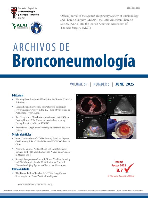
(A) Coronal CT image demonstrates a well-circumscribed oval nodule (arrow) within the left lower lobar bronchus, exhibiting smooth margins without lobulation. (B, C) Contrast-enhanced axial CT sections reveal progressive enhancement of the endobronchial lesion (arrow) during arterial and venous phases. (D) Histopathological examination shows tumor cells arranged in nested patterns, characterized by polygonal morphology with abundant eosinophilic cytoplasm, round-to-oval nuclei containing prominent nucleoli.
B.H.L. contributed to the acquisition and analysis of data; B.L.L. contributed to drafting the text and preparing the figures.
DeclarationNever used any artificial intelligence tools or technologies to assist in generating the paper.
FundJiangxi Provincial Administration of Traditional Chinese Medicine, Science and Technology Program [Project No. 2023B0479].
Conflict of InterestThe authors disclose no conflicts.


