


To analyze the clinical and radiological characteristics and features of pleural fluid (PF) in patients with tuberculous pleural effusion (TPE).
MethodsRetrospective analysis of TPEs treated in our clinic over the last 23 years.
ResultsWe included 320 patients with TPE (70% men; median age 33 years). Mycobacterium tuberculosis was identified in the sputum or PF of 36% of the patients by microscopic examination, solid and liquid media cultures, or nucleic acid amplification tests. The greatest percentage of positive microbiological findings were associated with human immunodeficiency virus (HIV) co-infection (OR: 3.27), and with the presence in PF of proteins <4g/dl (OR: 3.53), neutrophils >60% (OR: 3.23), and glucose <40mg/dl (OR: 3.17). Pleural adenosine deaminase <35U/l was associated with TPEs that occupied less than half of the hemithorax (OR: 6.36) and with PF lactate dehydrogenase levels <500U/l (OR: 8.09). Radiological pulmonary opacities (30%) were more common in TPE occupying less than half of the hemithorax (OR: 2.73), in bilateral TPE (OR: 4.48), and in older patients (OR: 1.02). Factors predicting mortality were: HIV co-infection (OR: 24), proteins in PF <5g/dl (OR: 10), and greater age (OR: 1.05).
ConclusionsPatients with TPE and HIV co-infection and those with lower concentrations of proteins in PF had higher rates of positive microbiological results and death. Moreover, older patients had more pulmonary opacities and a higher incidence of death.
Analizar las características clinicorradiológicas y del líquido pleural (LP) de los pacientes con derrame pleural tuberculoso (DPT).
MétodosAnálisis retrospectivo de los DPT atendidos en nuestro centro durante los últimos 23 años.
ResultadosSe estudiaron 320 pacientes con DPT (70% varones; mediana de edad 33 años). En el 36% de los casos se identificó Mycobacterium tuberculosis en esputo o LP mediante examen microscópico, cultivos en medios sólidos y líquidos, o amplificación de ácidos nucleicos. El mayor porcentaje de identificaciones microbiológicas se relacionó con una co-infección por el virus de la inmunodeficiencia humana (VIH) (OR: 3,27) y con la presencia en LP de unas proteínas <4g/dl (OR: 3,53), neutrófilos >60% (OR: 3,23) o glucosa <40mg/dl (OR: 3,17). Una adenosina desaminasa pleural <35U/l se asoció con DPT que ocupaban <1/2 del hemitórax (OR: 6,36) y con niveles de lactato deshidrogenasa <500U/l (OR: 8,09) en LP. Las opacidades pulmonares radiológicas (30%) fueron más comunes si el DPT no alcanzaba la mitad del hemitórax (OR: 2,73), era bilateral (OR: 4,48) o los pacientes tenían mayor edad (OR: 1,02). Los factores predictores de mortalidad fueron: una co-infección VIH (OR: 24), proteínas en LP <5g/dl (OR: 10) y una mayor edad (OR: 1,05).
ConclusionesLos pacientes con DPT co-infectados por VIH o que presentan concentraciones bajas de proteínas en LP tienen mayor frecuencia de aislamientos microbiológicos y fallecimientos. Asimismo, los pacientes de mayor edad muestran más opacidades pulmonares y mortalidad.
Tuberculosis (TB) is a frequent cause of pleural effusion (PE): in a series of 3077 consecutive patients undergoing diagnostic thoracentesis in our hospital, TB was the fourth most common etiology.1 However, in around 2 thirds of cases, a diagnosis of probable tuberculous pleural effusion (TPE) is made on basis of the biochemical characteristics of pleural fluid (PF) (lymphocytic exudate with adenosine deaminase [ADA] ≥35U/l) after other potential causes of PE have been ruled out.2 In Catalonia, the incidence of tuberculosis has fallen in recent years from 48.3/100000 inhabitants in 1994 to 14.4/100000 in 2015.3 Among the cases of exclusively extrapulmonary TB reported in 2015, pleural involvement was the second most common (25%) after lymphatic disease (42%).3
Microbiological tests performed to confirm the diagnosis of TPE are often negative. In 2 Spanish series, sputum and PF cultures in Löwenstein–Jensen medium showed a sensitivity of 41% and 36%, respectively.4,5 Nucleic acid amplification techniques or polymerase chain reaction (PCR) are fast and highly specific, but lack sufficient sensitivity. For example, in a meta-analysis of 24 studies, the sensitivity of an automated PCR technique (Xpert®-MTB/RIF) in PF was 23% in patients with probable TPE.6
The aim of this study, conducted in a large series of patients with TPE, was to analyze variables associated with: (1) a greater frequency of microbiological findings in PF or sputum; (2) pleural ADA <35U/l; (3) the presence of pulmonary opacities on chest X-ray, and (4) exitus.
Materials and MethodsIn 1994, a database was implemented in our hospital to prospectively include the clinical characteristics of all consecutive adult patients who underwent diagnostic thoracentesis, irrespective of the medical department in which they were treated. For this study, all TPEs included in this database between January 1994 and September 2017 were retrospectively analyzed. Our center is a referral hospital with 440 beds that provides health coverage to 450000 individuals. The study protocol was approved by the local ethics committee (CEIC-1868).
We collected demographic (age and sex) and clinical (presence and duration of fever, human immunodeficiency virus [HIV] serology, tuberculin test, death during treatment) variables, radiological findings (PE size, location and loculation, and pulmonary opacities on chest X-ray), microbiological results (auramine or Ziehl–Neelsen staining, solid and liquid media cultures, and PCR techniques in sputum samples and PF), and PF biochemistry (total and differential white blood cell count, red blood cell count, protein, glucose, lactate dehydrogenase [LDH], and ADA). When more than 1 thoracentesis was performed in the same patient, only the results of the first were considered.
Microbiological TestingSputum and PF samples underwent auramine or Ziehl–Neelsen staining, and were cultured for mycobacteria in solid (Löwenstein–Jensen) and liquid (Middlebrook 7H9 modified with OADC and PANTA supplements) media. PCR was performed on sputum or PF samples using manual techniques until September 2010. The Xpert® MTB/RIF technique was then used until March 2012,7 when the FluoroType MTB® method was introduced (Hain Lifescience).
Pleural Fluid AnalysisPF samples were collected in 2 sterile heparin tubes (5ml) for biochemical and microbiological analysis. Biochemical testing was performed immediately after thoracentesis in Hitachi analyzers (711 and 911, or Hitachi Modular DP, Roche Diagnostic, Mannheim, Germany) until August 2013, when an AU5800 Beckmann Coulter analyzer was introduced (Brea, CA, USA). Both techniques use standardized spectrophotometric methods. The upper limits of normal for serum LDH were 480 and 370U/l, using the first and second autoanalyzers, respectively. However, all LDH values were corrected with respect to the most recent method. pH was analyzed using an arterial blood gases analyzer and ADA was determined with automated kinetic methods. Until February 2012, the PF cell count was performed manually in a Thoma chamber, and later with automatic counters (Sysmex XN-2000 and Sysmex XN-350; Roche Diagnostics, Mannheim, Germany).
Radiological DataChest X-rays were reviewed by an expert in thoracic radiology (MP). TPEs were considered small if they occupied less than half of the hemithorax; large if they occupied half or more of the hemithorax, but without complete opacification of the hemithorax; and massive, in the case of complete opacification. Loculated TPE was defined as compartmentalized PE, showing a convex form facing the pulmonary parenchyma, or not located in a dependent lung field. Pulmonary opacity was defined as a pulmonary parenchymal lesion caused by interstitial or airspace disease, including cavitation or cystic spaces (cyst larger than 1cm within the pulmonary parenchyma) or residual fibrous lesions.8 Since only 30% of patients had undergone chest computed tomography, and these procedures were performed on equipment of differing quality during the study period, we decided not to include this examination.
Diagnostic CriteriaA diagnosis of TPE was classified as confirmed when tuberculosis bacilli were isolated from sputum, PF or pleural biopsy, or if granulomas were observed in the latter. The diagnosis was classified as probable in patients who met the following conditions: (a) clinical picture consistent with TPE; (b) pleural exudate with ADA levels ≥35U/l; (c) resolution of PE with TB treatment, and d) other potential causes of PE could be reasonably ruled out during clinical follow-up of at least one year. A positive microbiological result was defined as tuberculosis bacilli identified in a sample of PF or sputum by microscopy, culture, or PCR.
Statistical AnalysisQuantitative variables were expressed as median (25% and 75% quartiles), and qualitative variables as absolute numbers and proportions. Comparisons between groups for quantitative and qualitative variables were performed using the Mann–Whitney and Fisher tests, respectively. We used the area under the curve of diagnostic efficiency to choose the best cut-off point for continuous variables (neutrophils, proteins, LDH, and glucose in PF) that could predict a positive microbiological result or an ADA below the cut-off point of diagnosis of tuberculosis. Logistic regression models were developed to identify factors associated with the presence of microbiological isolates, pleural ADA <35U/l (cut-off point used in our center),9 pulmonary radiological opacities, and exitus. Variables that were significant in the bivariate analysis were included in the multivariate analyses, and the stepwise logistic regression analysis with the Wald sequential exclusion method was then applied. Statistical significance was set at a P value of <.05. All calculations were performed using the SPSS statistical package version 24.0 (Chicago, Illinois, USA).
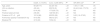
ResultsA total of 320 patients with TPE were analyzed, with a mean age of 33 (25–45) years, of whom 244 (70%) were men and 96 (30%) were women. Tuberculosis was classified as confirmed in 131 (41%) and probable in 189 (59%). Nine percent of patients had HIV co-infection and 78% had a positive tuberculin skin test (Table 1). Subjects with a positive tuberculin skin test had a significantly higher median age than those with negative results (37 vs 30 years, P=.03). Fever was recorded in 74% of cases over a median of 7 days (0–15) prior to thoracentesis. Fourteen patients died while receiving TB treatment, mostly in the first 2 months (64%).
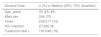
Main Characteristics of the Population With Tuberculous Pleural Effusion.
| General Data | n (%) or Median (25%–75% Quartiles) |
|---|---|
| Age, years | 33 (25–45) |
| Male sex | 244 (70) |
| Fever | 234/317 (74) |
| HIV infection | 27/290 (9) |
| Tuberculin test + | 191/246 (78) |
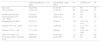
| Microbiological Data | 1st Sample | 2nd Sample (1st Negative) | 3rd Sample (1st and 2nd Negative) | Total |
|---|---|---|---|---|
| Auramine/ZN staining in sputum + | 26/219 (12) | 1/139 (0.7) | 3/92 (3) | 36/225 (16)a |
| Sputum culture + | 46/211 (22) | 8/115 (7) | 1/71 (1.4) | 66/228 (29)b |
| PCR in sputum + | 3/11 (27) | – | – | – |
| Auramine/ZN staining in PF + | 8/274 (3) | 0/12 | 8/274 (3) | |
| PF culture + | 51/296 (17) | 1/10 (10) | – | 52/296 (18) |
| PCR in PF + | 11/54 (20)c | – | – | – |
| Any isolate + | – | – | – | 115/320 (36) |
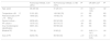
| Chest X-ray Data | |
|---|---|
| Site of PE | |
| Bilateral | 12/316 (4) |
| Right | 163/316 (51) |
| Left | 141/316 (45) |
| Size of PEd | |
| Small | 188/305 (62) |
| Large | 104/305 (34) |
| Massive | 13/305 (4) |
| Loculated PEd | 136/267 (51) |
| Pulmonary opacitiesd | |
| Ipsilateral | 58/81 (72) |
| Contralateral | 10/81 (12) |
| Bilateral | 13/81 (16) |
| Any site | 81/267 (30) |
| Progress | |
|---|---|
| Cure | 259/320 (81) |
| Active treatment | 15/320 (5) |
| Death | 14/320 (4) |
| Lost to follow-up | 32/320 (10) |
+: positive; HIV: human immunodeficiency virus; PCR: polymerase chain reaction; PE: pleural effusion; PF: pleural fluid; ZN: Ziehl–Neelsen stain.
Six patients had positive ZN staining but the order of the sample was unknown, so the result was only taken into account in the last column. If a patient's first sample was positive, the results of successive samples of the same specimen were not considered.
Microbiological confirmation was achieved in 115 (36%) patients in total (Table 1). In addition, 16 (84%) of 19 patients who underwent closed pleural biopsy were diagnosed by the demonstration of granulomas. Tuberculosis bacilli were isolated from 4 (57%) of the 7 pleural biopsies that were cultured. Sputum and PF cultures were positive in 29% and 18% of cases, respectively. Microbiological isolates were found more often in patients with HIV co-infection (56% vs 33%, P=.02), accounted for by high isolation rates in sputum cultures (50% vs 27%, P=.03), although not in PF (29% vs 17%, P=.13). Data on sensitivity to anti-TB drugs were available in only 23 patients: 7 had isolated resistance to pyrazinamide, and 1 was multiresistant (isoniazid, rifampicin, pyrazinamide, and ethambutol).
In a multivariate analysis (Table 2), factors associated with higher rates of microbiological identification were: 1 HIV co-infection (OR: 3.27), and PF containing proteins <4g/dl (OR: 3.53), neutrophils >60% (OR: 3.23), and glucose <40mg/dl (OR: 3.17).
Factors That Influence Positive Microbiological Identification in Tuberculous Pleural Effusion.
| Positive Identification, n=115 (36%) | Not Identified, n=205 (64%) | Pa | OR (95% CI)b | Pb | |
|---|---|---|---|---|---|
| Age, years | 36 (27–46) | 31 (25–44) | .03 | NS | NS |
| HIV co-infection | 15/102 (15) | 12/188 (6) | .02 | 3.27 (1.42–7.5) | .01 |
| Positive tuberculin test | 55/79 (70) | 136/167 (81) | .04 | NS | NS |
| Red blood cells in PF >10000/μl | 22/109 (20) | 20/203 (10) | .01 | NS | NS |
| Neutrophils in PF >60% | 18/107 (17) | 9/196 (5) | <.01 | 3.23 (1.28–8.14) | .01 |
| Glucose in PF <40mg/dl | 22/111 (20) | 12/205 (6) | <.01 | 3.17 (1.27–7.93) | .01 |
| Proteins in PF <4g/dl | 17/111 (15) | 9/205 (4) | <.01 | 3.53 (1.26–9.89) | .02 |
| LDH in PF >1000U/l | 44/107 (41) | 51/204 (25) | <.01 | NS | NS |
| Pulmonary opacity in standard X-ray | 36/95 (38) | 45/172 (26) | .05 | NS | NS |
Variables are expressed as median (quartiles 25–75) or n (%), as applicable.
HIV: human immunodeficiency virus; LDH: lactate dehydrogenase; NS: not significant; OR: odds ratio; PF: pleural fluid.
All TPE were exudates according to Light's criteria10; 10% (31/303) were predominantly neutrophilic (>50% neutrophils), while 53% (159/303) showed more than 90% lymphocytes on the white blood cell differential count. In 5 (36%) of 14 patients with neutrophilic TPE, a second thoracentesis showed predominantly lymphocytic effusion, although in all cases the absolute value and percentage of neutrophils in PF fell. Glucose in PF was <60mg/dl in 25%, and 9% had a pH of <7.20. PF contained >5g/dl proteins in 70% of cases. In 93% (294/315) of TPE, pleural ADA was ≥35U/l. Patients with ADA <35U/l tended to be older (36 vs 33 years), although the difference did not reach statistical significance.
In a multivariate analysis, pleural ADA <35U/l was associated with a TPE that occupied less than half the hemithorax and pleural LDH levels <500U/l (Table 3).
Factors Associated With Pleural ADA <35U/l in Tuberculous Pleural Effusion.
| ADA <35U/l, n=21 (7%) | ADA ≥35U/l, n=294 (93%) | Pa | OR: (95% CI)b | Pb | |
|---|---|---|---|---|---|
| Age, years | 36 (31–46) | 33 (25–45) | .18 | NS | NS |
| Glucose in PF >80mg/dl | 15 (71) | 132 (45) | .02 | NS | NS |
| Proteins in PF <5g/dl | 12 (57) | 81 (28) | <.01 | NS | NS |
| LDH in PF <500U/l | 15 (71) | 78 (27) | <.01 | 8.09 (2.69–24.35) | <.01 |
| pH in PF <7.40 | 14/18 (78) | 103/269 (38) | <.01 | NS | NS |
| Size of PE | 16/19 (84) | 168/282 (60) | .05 | 6.36 (1.38–29.3) | .02 |
Variables are expressed as median (quartiles 25–75) or n (%), as applicable.
ADA: adenosine deaminase; LDH: lactate dehydrogenase; NS: not significant; OR: odds ratio; PE: pleural effusion; PF: pleural fluid.
The most relevant radiological data are listed in Table 1. TPE was unilateral in 96% of cases, loculated in 51%, and occupied less than half of the hemithorax in 62% (a proportion that increased to 88% in patients with HIV co-infection). Among the 267 evaluable patients, pulmonary opacities were observed in 81 (30%), including residual fibrous lesions in 18, bronchogenic dissemination in 8, and cystic spaces in 4. Most of these opacities (68%) were located in the lung bases, while 17% were in the upper lobes, 5% in the right middle lobe, and 10% in multiple lobes. Mean age of patients with lesions in the upper lobes was similar to that of patients with lower lobe involvement (35 vs 38 years, P=.10).
As can be seen in Table 4, patients with pulmonary opacities were older (38 vs 31 years, P=.01), and had a larger percentage of positive sputum cultures (45% vs 20%, P<.01). However, there were no differences in the percentage of positive PF cultures (13% vs 17%, P=.44). Pulmonary opacities are associated with small (less than half the hemithorax) and bilateral PEs; these were the only variables that along with age were significant in a multivariate analysis.
Factors Associated With the Presence of a Pulmonary Opacity on Standard X-ray of the Chest of Patients With Tuberculous Pleural Effusion.
| Pulmonary Infiltrate, n=81 (30%) | No Pulmonary Infiltrate, n=186 (70%) | Pa | OR (95% CI)b | Pb | |
|---|---|---|---|---|---|
| Age, years | 38 (28–52) | 31 (25–41) | .01 | 1.02 (1–1.03) | .02 |
| Temperature >38°C | 51/81 (63) | 145/186 (78) | .01 | NS | NS |
| Red blood cells in PF >10000/μl | 18/80 (23) | 19/182 (10) | .01 | NS | NS |
| Sputum culture + | 26/58 (45) | 28/140 (20) | <.01 | NS | NS |
| PF culture + | 10/75 (13) | 30/174 (17) | .44 | NS | NS |
| Any microbiological identification | 36/81 (44) | 59/186 (32) | .05 | NS | NS |
| Bilateral PE | 7/81 (9) | 3/185 (2) | .01 | 4.48 (1.11–18.1) | .04 |
| Size of PE | 62/80 (78) | 96/182 (53) | <.01 | 2.73 (1.48–5.05) | .01 |
Variables are expressed as median (25%–75% quartile) or n (%), as applicable.
NS: not significant; OR: odds ratio; PE: pleural effusion; PF: pleural fluid.
Patients with loculated TPE more often had PF with glucose <60mg/dl (35% vs 20%, P=.01) or pH <7.20 (13% vs 4%, P=.01). However, the rate of microbiological isolates among these patients was not greater (36% vs 35%).
Prognostic DataPredictors of mortality (Table 5) were: HIV co-infection (OR: 24), proteins in PF <5g/dl (OR: 10) and an older age (OR: 1.05); this last variable was considered continuous. It should be noted that 3 of the 4 patients with HIV co-infection who died did so before the year 2000; the remaining subject died in 2013 after the simultaneous diagnosis of TPE and HIV infection, before antiretroviral therapy could be administered.
Predictive Factors of Death in Tuberculous Pleural Effusion.
| Death, n=14 (6%) | Cure, n=235 (94%) | Pa | OR (95% CI)b | Pb | |
|---|---|---|---|---|---|
| Age, years | 58 (39–77) | 32 (25–44) | <.01 | 1.05 (1–1.09) | .04 |
| HIV co-infection | 4/9 (44) | 20/218 (9) | <.01 | 24.44 (3.44–173.72) | <.01 |
| Fever | 6/14 (43) | 177/235 (75) | .01 | NS | NS |
| Positive tuberculin test | 2/8 (25) | 148/183 (81) | <.01 | NS | NS |
| Proteins in PF <5g/dl | 12/14 (86) | 65/233 (28) | <.01 | 9.63 (1.6–57.83) | .01 |
| Pulmonary opacity in standard X-ray | 9/13 (69) | 58/212 (27) | <.01 | NS | NS |
| Bilateral PE | 3/13 (23) | 7/234 (3) | <.01 | NS | NS |
Variables are expressed as median (quartiles 25–75) or n (%), as applicable.
HIV: human immunodeficiency virus; NS: not significant; OR, odds ratio; PE: pleural effusion; PF: pleural fluid.
This study describes the main clinical, microbiological, radiological and PF characteristics from a large series of patients with TPE, and determines the factors that are associated with a greater microbiological yield, lower levels of ADA in PF, the presence of pulmonary radiological opacities, and mortality.
It has been reported that the use of liquid culture media greatly increases the yield of mycobacterial isolates in TPE.11,12 However, despite the use of solid and liquid media, we only obtained an 18% positive culture rate in PF, a much lower figure than the 63% described by Ruan et al.,12 albeit similar to that of other studies.13 These discrepancies may be explained by factors such as the volume of PF processed (between 4 ml14 and 50ml,15 depending on the series), the type of liquid media used, the performance of a second culture,16 inoculation of PF in culture bottles at the bedside11,14,17,18 or in the laboratory, or the percentage of HIV co-infections (up to 80% in some studies).17 Specifically, in our series samples of only 5ml of PF were processed, with inoculation in specific culture media after the sample was received in the lab; serial PF culture were rarely performed (3%) and the population with HIV co-infection was significantly lower than in other studies.
In this study, factors associated with the isolation of Mycobacterium tuberculosis were HIV co-infection and PF containing proteins <4g/dl, neutrophils >60%, and glucose <40mg/dl. The greater frequency of mycobacterial isolates in patients with HIV co-infection is well known.17,19 In a series of 174 TPEs, PF cultures were positive in 64% of patients with HIV co-infection, compared to 30% of other cases.19 Immune system changes in this population probably favors a greater burden of bacilli in PF. Other series have investigated the relationship between biochemical and microbiological characteristics of PF in subjects with PTE.12,15,20 In this respect, a greater percentage of mycobacterial isolates has been associated with a predominance of neutrophils12,15,20 and low concentrations of glucose15,20 and proteins in PF.12 Neutrophils are predominant in the early stages of tuberculous pleural infection, before active immunity against the bacillus has developed.21 Low levels of glucose in PF have been associated with neutrophilic TPE22 (in our series, 45% of the neutrophilic TPEs had glucose levels <40mg/dl, compared to 6% in lymphocytic TPEs; data not shown), and are consequently associated with frequently positive cultures. Moreover, PTE with low proteins levels can indicate hypoproteinemia caused by malnutrition, also associated with altered immunity.23
With regard to the determination of pleural ADA, various cut-off points have been proposed to identify TPE, depending on the age of the patient.24,25 Tay and Tee24 proposed using an ADA of 72IU/l in patients aged ≤55 years and 26IU/l in patients aged >55 years to maintain a diagnostic sensitivity of 95% in both situations. The cut-off point of 55 years corresponded to the median age of the study population. However, in our series, only 15% of the subjects were older than 55 years. This fact may have influenced the lack of association between age and ADA values. The positive correlation between LDH and ADA in PF was described in a series of 80 TPEs (r=0.47, P<.001).24 Elevated LDH in PF indicates inflammation.26 A greater degree of inflammation probably increases the activation of lymphocytes and, consequently, the production of ADA.
TPE was accompanied by radiological pulmonary opacities in 30% of cases, a similar figure to that reported in previous studies (15%–27%).1 Paradoxically, in line with previous series,15,27 this finding was not associated with a greater frequency of positive sputum cultures in the multivariate analysis (in contrast to the univariate analysis). In pulmonary tuberculosis, the highest rate of positive sputum cultures is obtained in patients with opacities in the upper lobes or cavitations,28,29 manifestations that only were observed in 17% and 1.5% of patients in our study, respectively. In our series, more pulmonary opacities were observed in small TPEs, probably because it is easier to evaluate the lung parenchyma in these situations. The fact that a greater proportion of pulmonary opacities were found in bilateral PTEs may be because these may represent more disseminated disease.
TPE mortality was 6%, slightly lower than in other studies (7%–8%).20,30 Age and comorbidities (including HIV co-infection) were higher among subjects with TPE who died.20 However, in this study, HIV patients died primarily during the period before highly active antiretroviral therapy was fully developed, so the validity of this prognostic factor in TPE is questionable. In fact, in a prospective study of 196 patients with TPE conducted in Cameroon between 2007 and 2010, no prognostic differences were found between patients with or without HIV co-infection.31 On the other hand, low protein levels in PF may reflect hypoproteinemia and malnutrition, factors that carry a poor prognosis in pulmonary tuberculosis.32,33
The main limitation of this study is its retrospective design, with the consequent loss of data. For example, the tuberculin test was not used in 23% of cases; no sputum and PF cultures were performed in 29% and 8% of subjects, respectively, and PCR techniques were not used in 83%. Another potential limitation is the inclusion of a significant percentage of probable TPEs, although the criteria used to define them is widely accepted in the literature. This is rooted in the small number of closed pleural biopsies that are performed in our hospital, and the fact that most study subjects were referred from the Internal Medicine department. Finally, the associations identified between several variables may be valid only under the conditions of this study. Thus, not only has the prognosis for HIV infection improved substantially in present times, but the prevalence of pulmonary opacities in TPE is also much higher in CT studies than in standard X-ray,2,34 although CT is not routinely indicated in the setting of pleural tuberculosis.
In summary, factors such as HIV co-infection and low concentrations of proteins in PF are associated with a greater percentage of microbiological findings and a higher mortality rate. Moreover, older patients had more pulmonary opacities on radiological studies and a higher rate of death. Finally, smaller TPEs are accompanied by lower levels of pleural ADA and more pulmonary opacities. These results require confirmation in prospective studies.
Conflict of InterestsThe authors state that they have no conflict of interests.
Please cite this article as: Bielsa S, Acosta C, Pardina M, Civit C, Porcel JM. Derrame pleural tuberculoso: características clínicas de 320 pacientes. Arch Bronconeumol. 2019;55:17–22.


