
High-flow oxygen therapy (HFO) has gained popularity for the treatment of patients with acute respiratory failure, especially during the COVID-19 pandemic.
HFO is characterized for two main phenomenon which are an improvement of oxygenation and a reduction of the respiratory rate (RR). One of its well-known benefits is its capacity to generate a positive end expiratory pressure (PEEP) but, to date, published studies have inferred alveolar pressures from nasopharyngeal recordings1 or end expiratory lung volume measurements using acoustic bioimpedance.2 In essence, the actual pressure reached in the alveolus has never been measured, and this measurement is not trivial considering the complex fractal dichotomy of the lung geometry.3
Imagine initiating a high-flow device implementing maximum flows, you will experience some discomfort taking the air if this flow exceeds your natural peak flow rate and, what is more important, you will exhale through the nose against a flow that sometimes exceeds 60L/m being extremely difficult. If this is perceptible by any healthy subject, we wonder if it can affect our patients and increase their inspiratory and especially expiratory resistive load. Because if so, we know from the most basic physiology4 that any increase in resistance, especially expiratory resistance implies a reduction in respiratory rate (RR). What if the reduction in the RR observed in patients on HFO therapy does not respond to comfort but to an increase in mechanical load and with it the respiratory effort that could lead to self-induced lung injury (P-SILI)?
There is yet some recent evidence and it is urgent to be aware of this, since this therapy will most likely have to be modified.
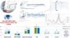
A recent prospective5 physiological study where a high-precision Millar® ultrathin pressure catheter6 able to reach the sixteenth bronchial generation of the Weibel Tree3 was guided with a bronchoscope and placed as distal as possible inside the patients’ lungs (Fig. 1), first under conventional oxygen therapy, and then under HFO 60L/m highlighted four relevant observations under HFO therapy: (1) intrapulmonary PEEP was demonstrated, reaching a mean of 7cmH2O, (2) inspiratory pressure decreased (PIP), (3) inspiratory time (IT) shortened and (4) expiratory time (ET) lengthened.
![Experimental setup and results (Ref. 5). High-flow oxygen therapy (HFO), respiratory rate (RR), positive end expiratory pressure (PEEP), self-induced lung injury (P-SILI), conventional oxygen therapy (COT), inspiratory pressure (PIP), inspiratory time (IT), expiratory time (ET), alveolar pressure gradient augmentation [PEEP-(-PIP)], heart rate (HR), mean invasive arterial pressure (MAP), arterial blood gases (ABG), arterial oxygen partial pressure (PaO2), arterial carbon dioxide partial pressure (PaCO2), bicarbonates (HCO3), base excess (BE) and oxygen saturation (SatO2).](https://static.elsevier.es/multimedia/03002896/0000005900000007/v3_202310232131/S0300289622005233/v3_202310232131/en/main.assets/gr1.jpeg?xkr=ue/ImdikoIMrsJoerZ+w9zfHQrK7O1GixshFj/RYvvhfIeFLIshOrwnoGDwJytw3P/oiFcT3h6qa0s2Be593HoRmdJ+4yy1McVp7/K4hrzIYldY6hCOhAB27JtMkOvV9uaE9wgmZuPeCSw5L8NmBdkUCXB0iSm6TGhRNTvgECWJSBSMEWV+TDxiP54CYIIJ9xiPz7Bjq7cHa1BPJGUtruAuqt9in4zbzAOWUfKGqsj3Fn5dHmOjNZ+3a4f4NIBqPeQDYvE9G2875ntELGWzmucAsQhfhnJ7t5c4EHGFOShc=)
Experimental setup and results (Ref. 5). High-flow oxygen therapy (HFO), respiratory rate (RR), positive end expiratory pressure (PEEP), self-induced lung injury (P-SILI), conventional oxygen therapy (COT), inspiratory pressure (PIP), inspiratory time (IT), expiratory time (ET), alveolar pressure gradient augmentation [PEEP-(-PIP)], heart rate (HR), mean invasive arterial pressure (MAP), arterial blood gases (ABG), arterial oxygen partial pressure (PaO2), arterial carbon dioxide partial pressure (PaCO2), bicarbonates (HCO3), base excess (BE) and oxygen saturation (SatO2).
In absence of specific measurements, the last two observations could indicate enhanced expiratory resistance. This resistance could lead to the lower RR proved in the registry, a finding firmly demonstrated when HFO was applied in healthy volunteers by Brochard and coworkers.7 In this paper, both inspiratory and expiratory resistance were measured accurately at different flow rates and increases significantly, reducing the RR, a fact that these authors warn should be taken into consideration in resistive patients (Asthma, COPD). Then, the reduction in RR at higher flows would not represent a mechanical improvement as we might believe, rather a mechanical overload assumed by the patient. Going back to the alveolar intrapulmonary pressure measurements,5 the demonstrated PEEP and the reduced PIP under extreme HFO, unleashes swings conditioned by alveolar pressure gradient augmentation [PEEP-(-PIP)] and this shearing may translate in alveolar stress and strain situation and thus a potential P-SILI8 that may be taking place while using such high flow rates in widely damaged lungs, as in acute respiratory distress syndrome (ARDS).
After considering these novel studies, two important questions must be addressed: Should we personalize HFO? and how can we do it? In clinical practice, the mechanical variables needed to adjust an optimal flow for each patient (transpulmonary pressure, resistances, alveolar pressure, …) are not available. Widely used indices9 to predict HFO failure are based on SatO2/FiO2 and RR. Clinicians could feel encouraged to rise HFO following the trend: the greater the flow, the better the oxygenation and the lower the RR. Even this is true, this RR reduction is probably due to a hidden resistive load able of course to generate PEEP but maybe at a too high workload cost. In any equation that seeks to determine the effectiveness of HFO the concept of respiratory effort should appear and be bedside monitored.
We would recommend setting up lower flows (HFO 30–40L/m) in resistive patients as some authors also have suggested previously10 given that higher flows seem not to improve the effort measured by esophageal pressure–time product per minute. Given that there is a clear commitment to HFO in combined treatment with non-invasive ventilation (NIV) in hypercapnic acute respiratory failure, these observations must be considered.
For lung damaged subjects as in ARDS, further studies must define this titration and verify the existence of an increase transpulmonary pressure at higher flows accompanying the demonstrated change in the alveolar ultrastructure pressure gradient5 able to generate the possible P-SILI mentioned above. We would recommend revising the Berlin definition11 by withdrawing definitively NIV from the treatment of mild ARDS in favor of “titrated” HFO and/or continuous positive airway pressure (CPAP) given the Florali study,12 in which the mortality of ARDS patients was higher under NIV than HFO, probably due to the fact that NIV cannot ensure minute ventilation for lung protection and therefore can degenerate into ventilator lung injury (VILI).13 But this appeal does not mean a support to a liberal flow strategy. On the contrary, we believe extreme HFO rates must be avoided given these novel physiological results5 where alveolar pressure swings can reach a mean of 13.66cmH2O at HFO 60L/m translating a possible P-SILI, worsening lung damage and probably delaying early intubation.
In a closer relation with COVID-19, CPAP or HFO should be proposed, as they have shown optimal results reducing the rate of intubation and mortality.14 However, just as NIV can induce VILI, it is probably imperative to title HFO to avoid P-SILI. This new concept must urgently be clarified since the RECOVERY trial15 did not detect better outcomes with HFO versus CPAP probably because of the known methodological limitations of the trial (timing of the study, selection of patients and arbitrarily in the choice of the device, as main problems) but we cannot rule out that these hidden phenomena may have influenced the results in an entity with an already very high respiratory drive. New larger physiological studies and probably new clinical trials are needed to conclude with certainty this important issue.
In conclusion we could really be facing a problem of titration or customization of the flow in HFO therapy in obstructive and ARDS COVID-19 or non-COVID-19 patients.


