


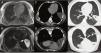
Our patient was a 39-year-old man who underwent echocardiography because of clinical palpitations. A mass was visualized in the left lateral-apical region and tentatively diagnosed as a pericardial cyst. Magnetic resonance imaging (MRI) was performed showing a fluid collection in the left fissure with a portion in the cardiophrenic angle (Fig. 1A and B).
A–F) Migrating pleuropericardial cyst: A) MRI T1-enhanced axial image showing hypointense lesion (asterisk) in the left fissure. B) MRI T2-enhanced axial image showing hyperintense lesion (asterisk) in left fissure extending to pericardium (arrow). C) CT axial image obtained in supine position revealing a lesion of fluid density (asterisk). D) CT axial image obtained in prone position showing a lesion of fluid density (asterisk) with anterior shift compared to in the supine position. E) CT axial image obtained in supine position in lung window showing lesion (asterisk) in.
We decided to expand the study with computed tomography (CT) focusing on the lesion, collecting images in supine and prone decubitus. A fluid collection was observed in the left fissure when the patient was supine, in the paracardiac region when he was in right lateral decubitus, and in the anterior region in prone decubitus (Fig. 1C and F). Part of the collection remained in the cardiophrenic angle in all scans, suggesting a diagnosis of migratory pleuropericardial cyst.
Pleuropericardial cyst is a rare entity that originates from an erroneous division of the coelom, usually in the right anterior cardiophrenic angle. If the cysts are pedunculated, they can move around the chest cavity, a phenomenon known as migrating pleuropericardial cyst. Diagnosis is obtained from imaging tests when a cystic chest mass is observed that is dependent on the pericardium and changes position with the patient’s movement, appearing in the lowest regions due to the force of gravity1,2.
Please cite this article as: Cabedo-Esteve L, Pérez-Serrano C, Vollmer I. Quiste pleuropericárdico migratorio: a propósito de un caso. Arch Bronconeumol. 2021;57:648.


