


We report the case of a 59-year-old woman with a pulmonary mass, who presented due to a 3-month history of discomfort in the left hemithorax. Chest computed tomography showed a mass of 67mm×87mm with defined borders, soft-tissue density, and peripheral calcifications, in the lower left lobe; and, in the same lobe, another lesion with the same characteristics, measuring 26mm×39mm.
Clinical staging was negative, and 15 days after diagnosis a left lower lobectomy was performed with resection extended to the diaphragm. The pathology report revealed 2 tumors, one 80mm×63mm and the other 45mm×34mm, with disease-free margins. Both tumors had the same microscopy report of spindle cell neoplasm with lengthened, hyperchromatic nuclei, and areas of epithelioid cells. Diaphragmatic muscle fibers showed no signs of malignant infiltration. Immunohistochemistry was positive for CD34 and Bcl-2, with Ki67 proliferation index of 2%. Diagnosis was intrapulmonary fibrous tumor with pleural involvement.
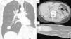
The patient remained asymptomatic and disease-free until 16 months after the intervention, when multiple nodules consistent with metastases were detected in the lungs, pleura, liver, bone, and muscles (Fig. 1).
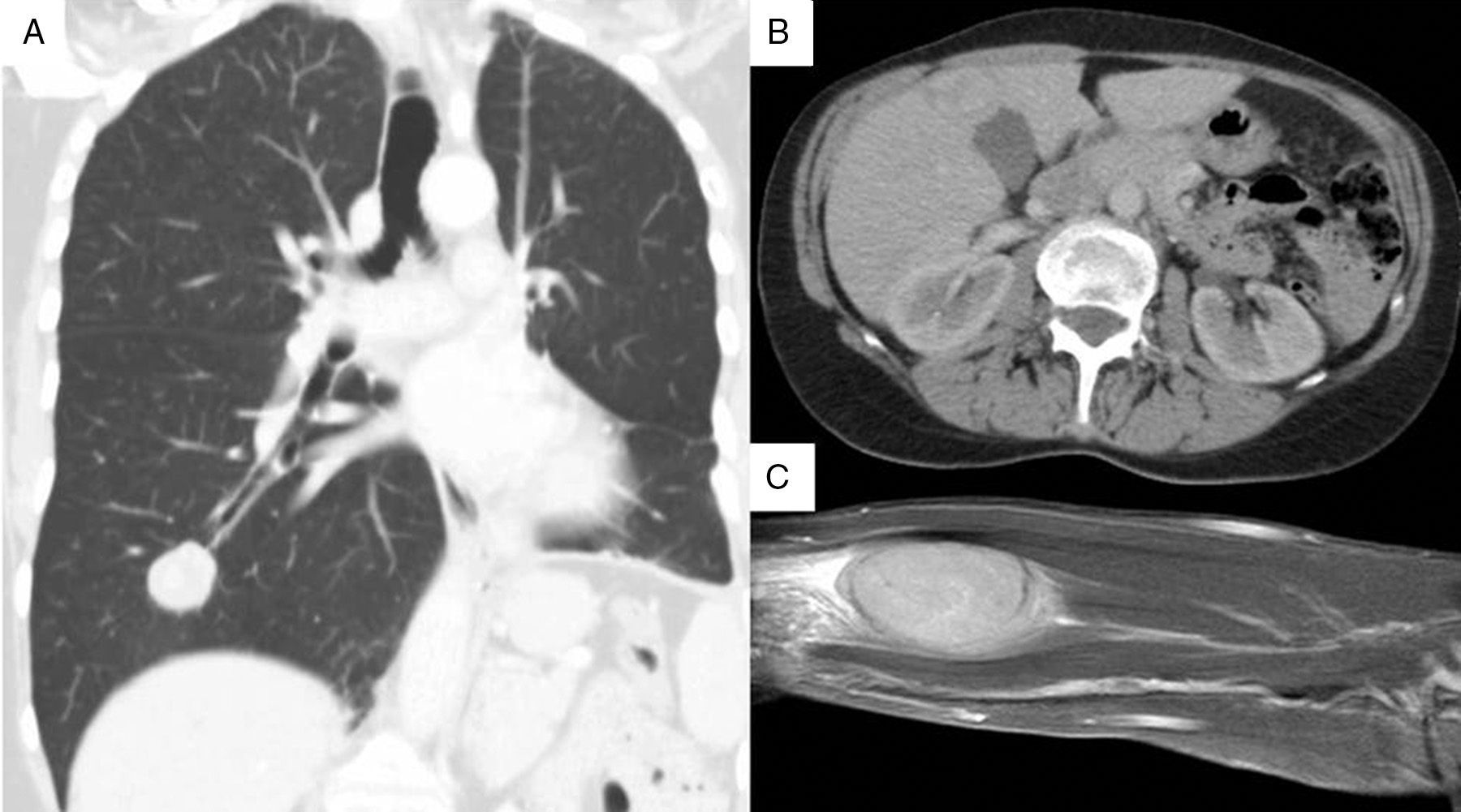
(A) Chest computed tomography, showing a 25-mm nodular image in the basal segment of the right lung field. (B) Tomography slice of the abdomen, showing a mass in liver segment V, measuring approximately 30mm. (C) Magnetic resonance image of nodular formation located between the muscle planes of the palm and distal third of the right forearm, 50mm×30mm×20mm.
A lung biopsy was performed and the pathology findings showed the same microscopic characteristics as the previous lesions, with a Ki67 proliferation index of 10%.
Metastatic fibrous tumor was diagnosed and the chemotherapy committee decided to administer 4 cycles of doxorubicin.
The disease progressed with tumor growth, pain, cholestasis, pyloric syndrome, and dyspnea. Subsequently, an incisional biopsy of a lesion on the patient's forearm revealed the same tumor characteristics, but this time Ki67 proliferation index was 25%.
Currently, 2 years after diagnosis, the patient is receiving second-line paclitaxel, with little response, and local radiation therapy.1,2
Intrapulmonary fibrous tumor forms from submesothelial fibroblasts in the pulmonary parenchyma, and is very rare.3 It is a slow growing tumor, with a benign course, and detection is incidental. Even more rare is the malignant form, which we present here.
Radiologically, it is visualized as a nodule with defined borders, with or without internal calcifications, and heterogeneous uptake of intravenous contrast medium.4 The histological features are those described above, and it is identical to pleural fibrous tumor. Immunohistochemistry is positive for CD34 antibody in 100% of cases, CD99 antibody in 70% of cases, Bcl-2, SMA and epithelial membrane antigen in 20% of cases, and vimentin in 90% of cases; it is negative for cytokeratins, actin, desmin, and S100 protein.3
Treatment of choice is resection with disease-free margins.4,5 Systemic chemotherapy may be administered, but doubts remain as to its usefulness.1,2
Five-year survival is greater than 90% and the rate of recurrence is 1.4% in benign forms and between 9% and 19% in malignant variants.3
As demonstrated by our patient, the biological behavior and the unpredictable course of fibrous tumor make long-term follow-up essential.
Please cite this article as: Schiavoni E, Padilla FA, Bustos M. Tumor fibroso solitario intrapulmonar: ¿benigno o maligno? Arch Bronconeumol. 2016;52:225–226.


