


La ecografía endobronquial (EBUS) es una nueva técnica que puede utilizarse para el diagnóstico por la imagen del tromboembolismo pulmonar (TEP) central. En 8 casos de nuestra clínica se utilizó la EBUS a causa de adenopatías mediastínicas o lesiones nodulares paramediastínicas, y al mismo tiempo se obtuvieron imágenes de un TEP mediante la EBUS. El TEP se diagnosticó antes de la EBUS mediante tomografía computarizada (TC) pulmonar en todos los casos (5 mujeres y 3 varones). Los defectos de repleción de todos los casos compatibles con un TEP se esclarecieron mediante angio-TC. La EBUS puede ser un método alternativo para el diagnóstico del TEP, ya que puede indicar la presencia de un trombo en arterias pulmonares centrales en casos en los que la hemodinámica es estable.
Endobronchial ultrasound (EBUS) is a new technique that can be used for the diagnostic imaging of central pulmonary thromboembolism (PE). In eight cases at our clinic, EBUS was used because of mediastinal lymphadenopathies or paramediastinal nodular lesions and at the same time images were obtained of a PE by means of EBUS. The PE was diagnosed before the EBUS with computed tomography (CT) of the lungs in all cases (5 women and 3 men). The repletion defects of all the cases compatible with a PE were clarified with CT-angiography. EBUS may be an alternative method for the diagnosis of PE, as it can indicate the presence of a thrombus in the central pulmonary arteries in hemodynamically-stable cases.
El tromboembolismo pulmonar (TEP) es un trastorno clínico relacionado con una oclusión súbita de la arteria pulmonar. Tiene una alta tasa de mortalidad y morbilidad. El TEP puede diagnosticarse erróneamente en los pacientes con comorbilidades. El empleo sistemático de angio-TC de tórax multicorte es la técnica de primera elección para el diagnóstico del TEP. El empleo de soluciones de contraste y la irradiación son sus principales limitaciones en los casos de insuficiencia renal y en el embarazo1,2. Por consiguiente, en estas distintas situaciones son esenciales otras pruebas diagnósticas (figs. 1 y 2).
 Imágenes de una embolia en la arteria pulmonar derecha en la EBUS.")
 Embolia bilateral en la arteria pulmonar central en la TC de tórax.")
La ecografía endobronquial (EBUS, endobronchial ultrasound) es una nueva técnica diagnóstica en la medicina pulmonar. Mediante el empleo de ultrasonidos en la punta del broncoscopio puede evaluarse el tejido de alrededor del sistema respiratorio y pueden aplicarse técnicas invasivas. Las principales indicaciones de la EBUS son las adenopatías mediastínicas y las masas centrales. Su empleo permite realizar con facilidad tanto el diagnóstico histopatológico de la masa (enfermedad maligna, tuberculosis, sarcoidosis, etc.) como la determinación del estadio de la enfermedad maligna3 (tablas 1 y 2).
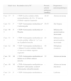
Características de los pacientes
| Edad | Sexo | Resultados de la TC | Presión arterial pulmonar (mmHg) | Diagnóstico anatomopatológico | |
| Caso 1 | 37 | F | • TEP• Lesión nodular sólida paramediastínica de 24×16 mm en el lóbulo superior izquierdo | 40-45 | Adenocarcinoma |
| Caso 2 | 61 | F | • TEP• Adenopatías mediastínicas• Cardiomegalia | 50-55 | Sin biopsia |
| Caso 3 | 50 | F | • TEP• Adenopatías mediastínicas• Pleuritis | – | Inflamación granulomatosa necrosante |
| Caso 4 | 51 | F | • TEP• Adenopatías mediastínicas• Lesión nodular sólida de 1×2 cm en el pulmón derecho | 45 | Inflamación granulomatosa necrosante |
| Caso 5 | 82 | M | • TEP• Adenopatías mediastínicas+hiliares• Lesión nodular sólida de 13mm en el lóbulo superior izquierdo | 40 | Metaplasia escamosa |
| Caso 6 | 50 | M | • TEP• Adenopatías mediastínicas• Infarto en el lóbulo inferior derecho | 35 | Adenocarcinoma |
| Caso 7 | 49 | M | • TEP• Adenopatías mediastínicas• Pleuritis• Adenopatías axilares• Adenopatías intraabdominales | – | Adenocarcinoma mucinoso |
| Caso 8 | 24 | F | • TEP• Adenopatías mediastínicas | – | Linfoma |
Identificación de trombos mediante TC y mediante EBUS
| Localización de los trombos en la TC | Localización de los trombos en la EBUS | |
| Caso 1 | AP central bilateral | AP central bilateral |
| Caso 2 | AP central bilateral | AP central bilateral |
| Caso 3 | Arteria del lóbulo inferior izquierdo | Arteria del lóbulo inferior izquierdo |
| Caso 4 | Arteria del lóbulo superior derecho | Arteria del lóbulo superior derecho |
| Caso 5 | AP principal izquierda | AP principal izquierda |
| Caso 6 | AP central bilateral | AP central bilateral |
| Caso 7 | AP central bilateral | AP central bilateral |
| Caso 8 | AP principal izquierda | AP principal izquierda |
AP: arteria pulmonar.
Recientemente se han publicado algunos artículos relativos al uso de la EBUS como método diagnóstico para el TEP central4-6. En este trabajo hemos evaluado el TEP en 8 pacientes mediante EBUS y hemos valorado la importancia de esta técnica en el diagnóstico del TEP.
Observaciones clínicasCaso 1Una mujer de 37 años de edad acudió al servicio de urgencias con tos y hemoptisis. En la TC de tórax se observó una lesión sólida nodular de 24×16mm, paramediastínica, en el lóbulo superior izquierdo y la presencia bilateral de un trombo en las arterias pulmonares. Se utilizó EBUS-FNA para el diagnóstico de la lesión nodular sólida y se observó un TEP en las arterias pulmonares centrales bilaterales.
Caso 2Una mujer de 61 años fue operada por una lesión de menisco. Después de la operación, la paciente sufrió un embolismo pulmonar masivo y se aplicó un tratamiento trombolítico. En la TC de tórax se observaron, además, ganglios linfáticos mediastínicos. Seis días después del tratamiento trombolítico la paciente estaba hemodinámicamente estable y se utilizó la EBUS para el examen de los ganglios linfáticos. Dado que las imágenes de los ganglios tenían unas características ecográficas benignas, no se aplicó la aspiración con aguja fina (FNA, fine-needle aspiration). Durante la EBUS se observó la presencia de trombos arteriales pulmonares bilaterales.
Caso 3Una mujer de 50 años acudió al servicio de urgencias por la aparición súbita de disnea y fatiga. En la TC de tórax se observó la presencia de un trombo en la arteria del lóbulo inferior izquierdo, así como ganglios linfáticos de 2cm mediastínicos, hiliares y subcarinales. Se utilizó una EBUS-FNA de los ganglios, y durante la EBUS se visualizó el trombo en la arterial del lóbulo inferior izquierdo. En el examen anatomopatológico se diagnosticó una necrosis caseosa.
Caso 4Una mujer de 51 años acudió al hospital por disnea y dolor torácico. En la TC de tórax se observó la presencia de una lesión nodular sólida de 2cm en el pulmón derecho, adenopatías mediastínicas y un TEP en la arteria del lóbulo superior derecho. Se utilizó una EBUS-FNA de la lesión y los ganglios linfáticos, y durante la EBUS se visualizó el trombo en la arteria del lóbulo superior derecho. En el examen anatomopatológico se identificó una inflamación granulomatosa necrosante.
Caso 5Un varón de 82 años acudió al hospital por disnea y dolor torácico. En la TC de tórax se observó una lesión nodular sólida de 13mm en el lóbulo superior izquierdo, junto con adenopatías mediastínicas e hiliares y un TEP en la arteria pulmonar principal izquierda. Se utilizó una EBUS-FNA de la lesión y los ganglios linfáticos, y durante la EBUS se visualizó el trombo en la arteria pulmonar principal izquierda. En el examen patológico se observó una metaplasia escamosa.
Caso 6Un varón de 50 años acudió al servicio de urgencias por hemoptisis y dolor torácico. En la TC de tórax se observó un infarto en el lóbulo inferior derecho, junto con adenopatías mediastínicas y un TEP bilateral en las arterias pulmonares centrales. Se utilizó una EBUS-FNA de los ganglios linfáticos, y durante la EBUS se visualizó un trombo en las arterias pulmonares centrales bilaterales. El examen patológico indicó la presencia de un adenocarcinoma.
Caso 7Un varón de 49 años acudió al servicio de urgencias por hemoptisis, dolor torácico y edema en el cuello. En la TC de tórax se observaron adenopatías mediastínicas, axilares, una pleuritis mínima bilateral y un TEP en las arterias pulmonares centrales bilaterales. Se utilizó una EBUS-FNA de los ganglios linfáticos, y durante la EBUS se visualizó un trombo en las arterias pulmonares centrales bilaterales. En el examen patológico se observó un adenocarcinoma mucinoso.
Caso 8Una mujer de 24 años acudió al hospital por síntomas de tos y dolor torácico de un mes de evolución. En la TC de tórax se observaron múltiples ganglios linfáticos y un trombo en la vena cava superior. Para el diagnóstico se utilizó una EBUS-FNA, y durante el examen de EBUS, además del trombo en la vena cava superior, se observó la presencia de un trombo en la arteria pulmonar superior derecha. El examen patológico de las biopsias de los ganglios linfáticos indicó la presencia de un linfoma de células B.
DiscusiónEl TEP es un trastorno pulmonar frecuente y difícil de diagnosticar que constituye la principal urgencia pulmonar con peligro para la vida del paciente. Generalmente los pacientes presentan un dolor torácico pleurítico súbito con dificultad respiratoria, pero a veces los pacientes con TEP tienen síntomas inespecíficos que pueden hacer que el diagnóstico de TEP se retrase o no llegue a establecerse. Este retraso en el diagnóstico es el factor más importante de mortalidad del TEP. Por consiguiente, los pacientes en los que se sospecha la presencia de un TEP deben ser evaluados y diagnosticados con rapidez. No existe ninguna prueba diagnóstica no invasiva que sea exacta1,2. Recientemente la angio-TC multicorte de tórax ha pasado a ser el método de primera elección para el diagnóstico del TEP. Con el aumento del número de detectores pueden visualizarse con facilidad los trombos periféricos7. Sin embargo, la dosis de radiación aumenta con el número de detectores, el área de examen y el grosor del corte. Además, para la angiografía es preciso utilizar una solución de contraste yodada que es nefrotóxica. Por consiguiente, en las mujeres embarazadas, en los pacientes con insuficiencia renal y en los pacientes alérgicos al contraste la angio-TC no es un método apropiado y deben usarse otras pruebas diagnósticas para establecer la presencia de un TEP. Según el estudio PIOPED II, la angio-TC no puede utilizarse en el 24% de los pacientes con TEP a causa de las contraindicaciones2. La gammagrafía de ventilación-perfusión es otra prueba diagnóstica utilizada para el TEP. Para esta exploración deben utilizarse también isótopos radiactivos. La gammagrafía V/Q es apropiada en los pacientes en los que se sospecha un TEP y que presentan radiografía de tórax normal y características cardiopulmonares básicas normales. Si hay una radiografía de tórax anormal, el valor diagnóstico de la gammagrafía se reduce8. La resonancia magnética puede utilizarse también para visualizar el trombo en la arteria pulmonar. Sin embargo, los artefactos producidos por el movimiento, la alergia al gadolinio y la inaplicabilidad en situaciones de urgencia constituyen sus principales inconvenientes9. En aproximadamente el 70% de los pacientes con TEP hay una trombosis venosa profunda (TVP). El diagnóstico de la TVP constituye un dato importante para iniciar un tratamiento anticoagulante en los pacientes en los que se sospecha un TEP. Sin embargo, el hecho de descartar la TVP no descarta la presencia de un TEP. No existe una prueba diagnóstica definitiva, con complicaciones mínimas, que pueda aplicarse en todos los casos. En consecuencia, es preciso utilizar muchas pruebas diagnósticas de forma combinada1,2.
La EBUS se emplea generalmente para la biopsia de las adenopatías mediastínicas e hiliares o las masas. Las arterias pulmonares son del orden de 5mm en la zona traqueobronquial. Por consiguiente, la EBUS puede visualizarlas con facilidad. Nosotros utilizamos una sonda de ecografía convexa (Olympus EBUS-CV-180, Tokio, Japón) para la evaluación broncoscópica. El diámetro externo de la CP-EBUS es de 6,9mm. Dispone de un canal de trabajo de 2,0mm y de una óptica de visión anterógrada oblicua de 30grados. La penetración máxima del transductor ecográfico lineal es de 50mm, y está conectado a un procesador que permite visualizar el flujo de los vasos mediastínicos e hiliares10.Casoni et al. utilizaron la EBUS en un paciente que presentaba una lesión intraarterial de baja densidad sospechosa en la angiografía pulmonar y publicaron por primera vez el diagnóstico de un TEP4. Aumiller et al. diagnosticaron de forma incidental un TEP en el examen de determinación del estadio de 3 pacientes con cáncer de pulmón mediante EBUS. Posteriormente se inició un estudio multicéntrico para evaluar la presencia de TEP mediante CP-EBUS en las primeras 24h siguientes al diagnóstico del TEP mediante angio-TC. Se detectaron con facilidad los trombos en las arterias pulmonares en 28 de 32 pacientes. Un total de 97 de los 101 trombos diagnosticados mediante angio-TC fueron confirmados con la EBUS (sensibilidad del 96%). La EBUS permite visualizar el tronco pulmonar, las arterias pulmonares principales y las arterias lobulares. Uno de los 4 trombos que no pudieron visualizarse se encontraba en la arteria del lóbulo medio y 3 estaban en la arteria del lóbulo superior izquierdo. Durante el estudio, ninguno de los pacientes presentó complicación alguna. En consecuencia, los autores llegaron a la conclusión de que la EBUS era una prueba diagnóstica fiable para el TEP de localización central5. Después de este estudio, se han publicado múltiples descripciones de casos de uso de la EBUS en el diagnóstico del TEP6,11-13.
En el presente estudio, todos los pacientes estaban hemodinámicamente estables y fueron sedados durante la broncoscopia. La EBUS se llevó a cabo utilizando un abordaje oral, con anestesia consciente en todos los pacientes. Se mantuvo un registro constante de la presión arterial, la frecuencia del pulso y la saturación de oxígeno durante todos los procesos. Las arterias pulmonares principales se visualizaron tras la evaluación de las adenopatías mediastínicas en todos los casos. El broncoscopista sabía dónde estaban los defectos de repleción observados en las arterias pulmonares en la TC. Las arterias pulmonares permeables no muestran ecos. En nuestros casos, cuando se visualizaron zonas ecogénicas, el broncoscopista confirmó la presencia del trombo con el empleo del modo Doppler.
En conclusión, hemos evaluado el TEP mediante EBUS de manera fácil y fiable. En las guías del TEP no se hace referencia a la EBUS en relación con el diagnóstico del TEP. Dado que la angio-TC no pudo aplicarse en un número sustancial de pacientes, el diagnóstico por la imagen de los trombos bajo guía de EBUS puede ser una buena alternativa en los pacientes hemodinámicamente estables para el diagnóstico del TEP. Además, en las unidades de cuidados intensivos el traslado de algunos pacientes a la unidad de TC constituye un problema importante, y la EBUS puede ser útil en estos casos para el diagnóstico del TEP. Por otra parte, la EBUS puede visualizar la vascularidad central y permite diferenciar con facilidad las enfermedades malignas vasculares del TEP. Sin embargo, serán necesarios más estudios controlados, aleatorizados y de diseño ciego simple, dado que en nuestro trabajo, y también en el estudio de Aumiller et al., los broncoscopistas conocían la localización del TEP en la angio-TC5.
Conflicto de interesesLos autores declaran no tener ningún conflicto de intereses.


