


Surgical removal of intrathoracic goiter can be performed by a cervical approach in the majority of patients. Review of literature shows that experienced surgeons need to perform an extracervical approach in 2–3% of cases. In spite of surgical management of substernal goiter is well defined, there is little available information about surgical approach of intrathoracic goiters extending beyond the aortic arch into the posterior mediastinum. We report two cases and propose combination of cervical incision and muscle-sparing lateral thoracotomy for posterior mediastinal goiter removal. In such cases, we do not favor sternotomy as posterior mediastinum is inaccessible due to the presence of heart and great vessels anterior to the thyroidal mass that would lead to perform a perilous blind dissection. Based on our experience, transcervical and thoracotomy approach is indicated for a complete and safe posterior mediastinal goiter removal.
La extirpación quirúrgica del bocio intratorácico puede realizarse a través de un abordaje cervical en la mayoría de los pacientes. La revisión de la literatura pone de manifiesto que los cirujanos experimentados precisan un abordaje extracervical en el 2-3% de los casos. A pesar de que el tratamiento quirúrgico del bocio retroesternal está bien definido, existe poca información acerca del abordaje quirúrgico de los bocios intratorácicos que se extienden más allá del cayado aórtico hacia el mediastino posterior. Presentamos 2 casos y proponemos una combinación de incisión cervical y toracotomía lateral con preservación muscular para la resección del bocio en el mediastino posterior. En este tipo de casos descartamos el uso de la esternotomía puesto que el mediastino posterior resulta inaccesible debido a la presencia del corazón y grandes vasos por delante de la masa tiroidea, lo cual podría llevar a realizar una peligrosa disección a ciegas. Según nuestra experiencia el abordaje transcervical combinado con la toracotomía está indicado para una resección completa y segura del bocio situado en el mediastino posterior.
A review of the literature confirms that posterior mediastinal goiter is rare, accounting for 10%–15% of all intrathoracic goiters (IG).1 While most IGs can be removed through a cervical incision, those extending into the posterior mediastinum may require additional extracervical incisions.2
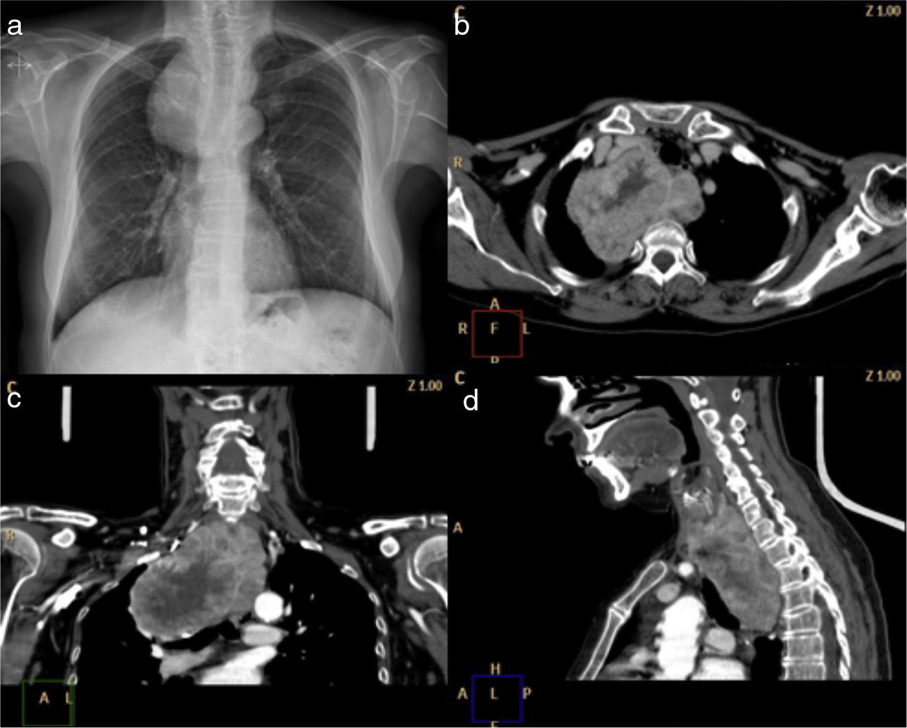
Case 1A 61-year-old woman was referred to our center with a 12-month history of subclinical hyperthyroidism. She had undergone subtotal thyroidectomy for a multinodular goiter seven years previously in another center. Thyroid function tests revealed hyperthyroidism. Computed tomography (CT) showed the retrotracheal cervical thyroid in continuity with the posterior mediastinal mass that extended beyond the aortic arch and the azygos vein (Fig. 1). The goiter was causing left shift of the trachea and the esophagus, terminating at the level of the left atrium, below the carina tracheae. Examination of the larynx excluded recurrent nerve paralysis. Spirometry results showed forced vital capacity (FVC) of 3.62 l (109%) and forced expiratory volume in 1 second (FEV1) of 2.88 l (114%).
Right hemithyroidectomy was scheduled. In the first stage, a cervical approach was used for resection of the residual right thyroid. Extension of the thyroid tissue behind the trachea toward the left side was observed. The mass crossed the mid-line and extended toward the superior thoracic outlet. All surrounding tissue adhesions were removed from the cervical region and the mediastinal section of the thyroid was partially freed with gentle traction, until the superior thoracic inlet was reached. The cervical incision was then packed with gauze and the patient was turned on her left side and a right upper thoracotomy was performed. Resection of the mediastinal section of the thyroid gland was laborious due to the lack of space between the mass, the superior vena cava, the subclavian vein and the brachiocephalic artery. During surgery, a thyroid ima artery originating in the subclavian artery supplying the thyroid was identified. The mass was fully released with gentle traction via the thoracic incision. The pathology report was compatible with multinodular goiter. Progress after surgery was uneventful and the patient was discharged after one week.
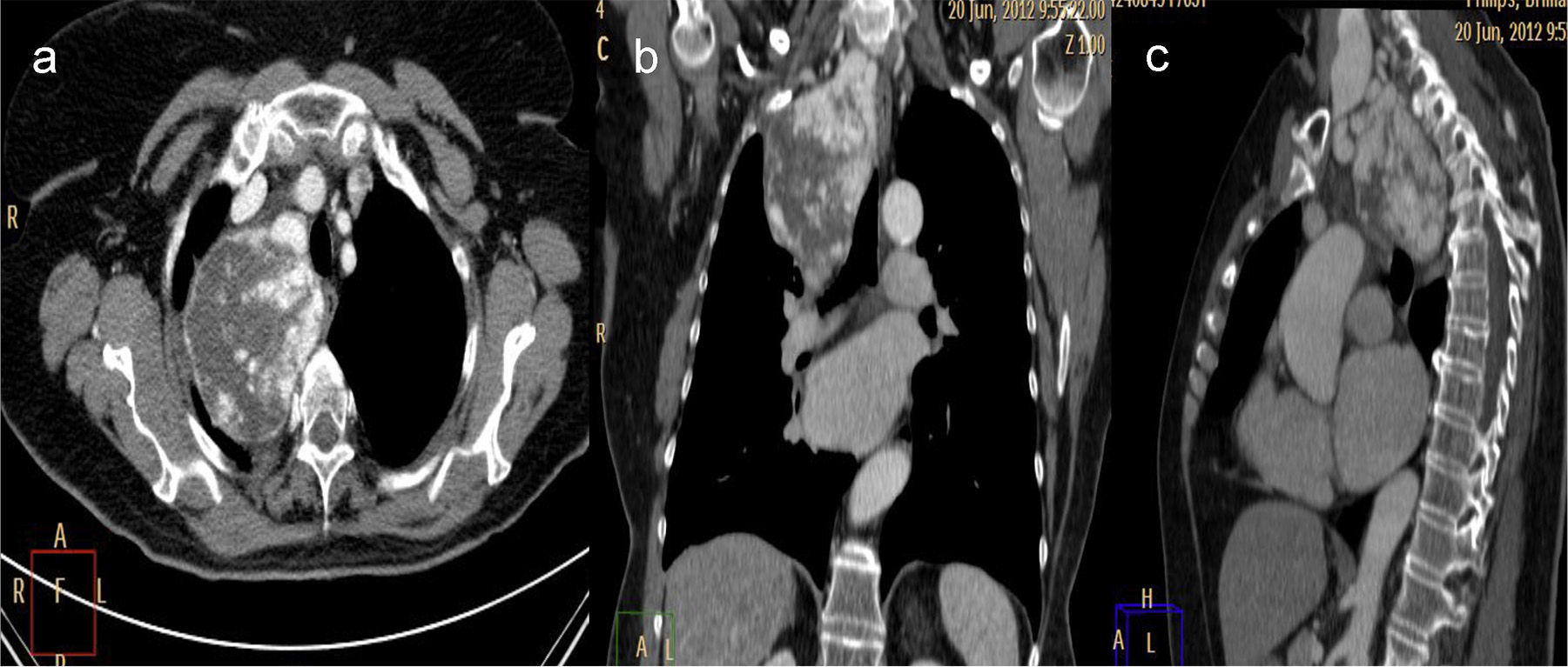
Case 2A 64-year-old woman was admitted to our center due to a 6-month history of IG. The IG had been identified during a routine evaluation of atrial fibrillation. Thyroid function tests were normal. CT revealed a lobulated mass in the thyroid gland adjacent to the lower pole of the right thyroid lobe (Fig. 2). The mass was seen to extend in a retrotracheal direction to a distance of 3cm below the carina tracheae. Spirometry results showed an FVC of 2.63 l (60%) and an FEV1 of 1.92 l (62%).
A cervical approach with lateral thoracotomy for right hemithyroidectomy was selected. First, a cervical Kocher incision was made. The thyroid mass extended toward the left, crossing the mid-line and descending along the superior thoracic inlet. Careful attention was paid to the right laryngeal nerve and to the inferior mesenteric arterial blood supply during dissection of the mass in the thoracic outlet. All adhesions in the right thyroid lobe were removed from the cervical region and dissection continued to above the trachea. The Kocher incision was sutured, and the patient was turned on her left side. The procedure continued with a right lateral thoracotomy. The mediastinal section of the right thyroid gland terminated 3cm below the carina tracheae. The mediastinal pleura was opened and the thyroid mass was completely removed. Pathology testing revealed multinodular goiter. Transient dysphonia occurred during the postoperative period. The patient was discharged after one week.
DiscussionThe elective removal of IG is indicated when the diagnosis and extension of the mass have been determined. There is little information3,4 available on the surgical approach for retrotracheal IGs. The largest series published comprises 11 cases.5 Based on our experience, we propose the combination of an initial cervical incision followed by a right muscle-sparing lateral thoracotomy. The most important criteria for selecting patients who require cervical incision and thoracotomy are based on the characteristics observed on CT, thyroid volume and extension below the carina tracheae.6 Precise cervical dissection is essential for removal of all tissue adhesions from around the thyroid gland. Digital dissection following the lower thyroid lobe to the level of the thoracic outlet assists in mobilization. With this maneuver, the goiter can be withdrawn and extracted via the thoracotomy after sufficient removal of the posterior mediastinal surface. A high thoracotomy incision provides a wide operating field and allows monitoring and direct visualization of the large vessels and the posterior mediastinum. We do not recommend limiting the approach to a cervical incision due to the increased risk of uncontrollable bleeding, damage to the recurrent laryngeal nerve and incomplete resection of the goiter. We would also avoid sternotomy in these cases, since the heart and the large vessels immediately anterior to the thyroid mass make the posterior mediastinum inaccessible. In our opinion, in this situation the surgeon should not hesitate to carry out a muscle-sparing high lateral thoracotomy to achieve complete removal of the thyroid tissue while reducing intraoperative risk, particularly bleeding. Video-assisted thoracoscopy has been used for resection of the retrosternal goiter; however, we currently avoid it, due to the lack of evidence supporting its application in retrotracheal goiter surgery.
ConclusionThe combination of a cervical incision and muscle-sparing lateral thoracotomy provides a wide operating field and facilitates the safe and complete removal of posterior mediastinal goiter.
Conflict of InterestsThe authors declare no conflict of interests.
Please cite this article as: Ojanguren Arranz A, Baena Fustegueras JA, Ros López S, Santamaría Gómez M, Ojanguren Arranz I, Olsina Kissle JJ. Abordaje del bocio endotorácico en mediastino posterior: incisión transcervical y toracotomía lateral. Arch Bronconeumol. 2014;50:255–257


