


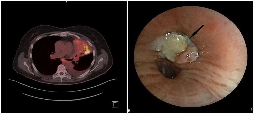
A 60-year-old woman was referred to the Pneumology Department due to suspicion of lung cancer. The patient presented with progressive dyspnea, intense asthenia and loss of 10kg of weight in less than three months. Chest X-ray and computed tomography both showed an obstructive lesion at the left upper lobe bronchus suggestive of endobronchial neoplasia. Positron emission tomography (Fig. 1, A) showed increased uptake of 18F-fluorodeoxyglucose from the lesion (SUVmáx 12.6). Flexible bronchoscopy revealed an endobronchial lesion that obstructed the entire left upper bronchus (Fig. 1, B) biopsies of said lesion showed abundant Actynomices colonies.
 hypemetabolic consolidation occupying the entire left upper lobe with obstruction of the upper lobe bronchus close to the bifurcation of the main bronchus; (B) flexible bronchoscopy shows an endobronchial lesion obstructing the left upper bronchus entrance (arrow).")
Actinomycosis is a bacterial infection caused by microorganisms of the genus Actynomices. Lung involvement is very rare, being the most frequent presentation the mandibular abscess.1 For its diagnosis, microbiological or anatomopathological identification of the microorganism is necessary. The treatment of choice is Penicillin between 6 and 12 months.1,2
FundingThere was no funding source in this study.
Conflict of interestThe authors declare that they have no conflict of interest to the publication of this article.


