


Accessory cardiac bronchus (ACB) is a rare congenital tracheobronchial branching abnormality with an incidence of 0.07–0.5% which originates from the medial wall of the intermediate or main bronchus.1–3
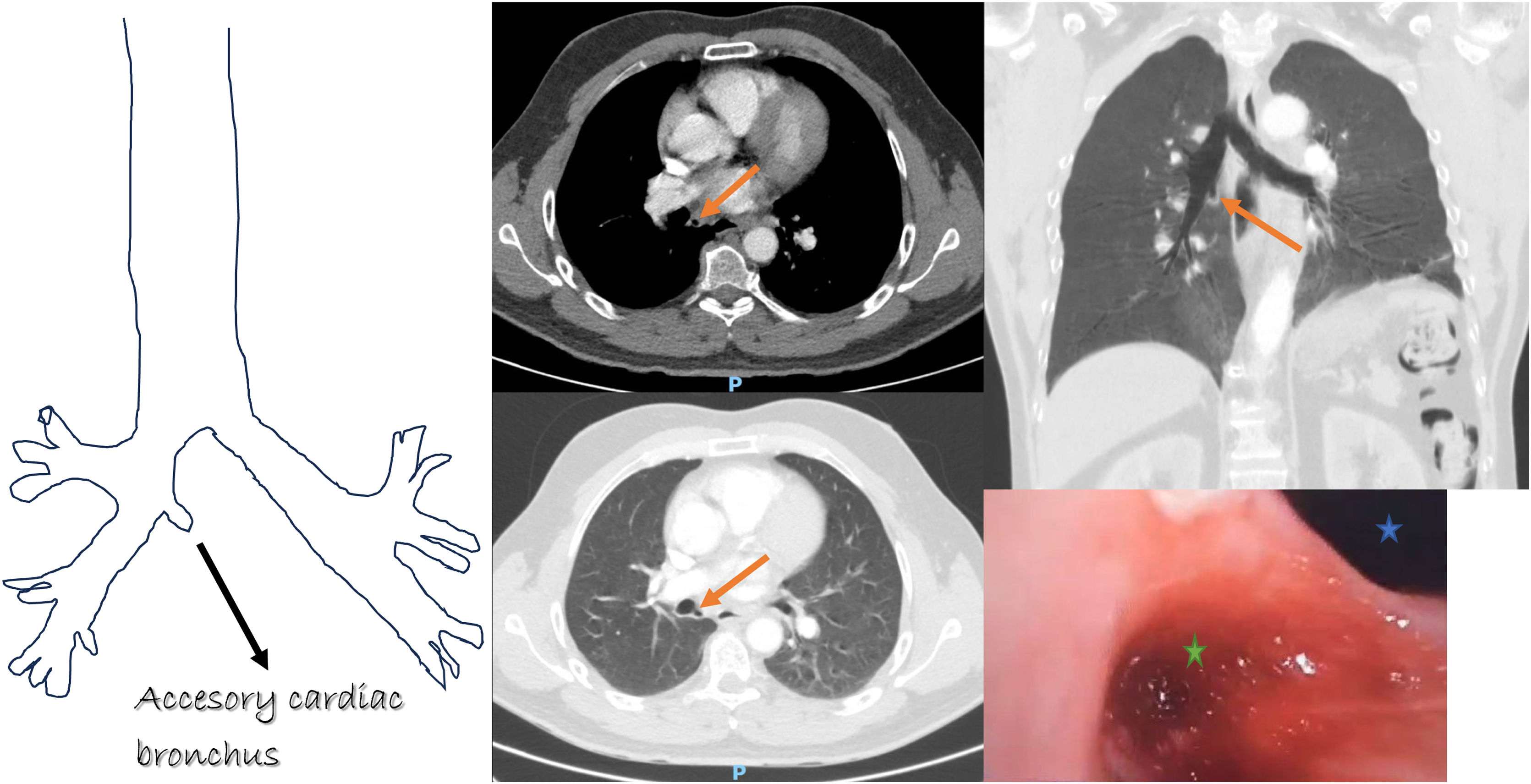
We present a 51-year-old male, an active smoker with a cumulative smoking rate of 18 pack-years and only for medical antecedent dorsal inguinal hernia being monitored by the pain unit. The patient had cough and whitish expectoration for 4 months before going to the emergency department due to hemoptysis of 150cc in about 10h with a previous episode 8 days before at home of hemoptysis of about 200cc in 24h. The physical examination and different blood test were normal. The chest X-ray revealed a slight right paracardiac infiltrate, and chest computed tomography highlighted the presence of a ACB that originated at the entrance to the bronchus intermedius on its medial wall (Fig. 1), approximately 10mm deep, and with an inflammatory halo on the wall of the sac. Fiberoptic bronchoscopy was performed, showing a tracheal bronchus and a complementary right segment 6 bronchus and an orifice at the entrance of the intermediate bronchus in its medial wall (ACB) from which fresh blood emanated (Fig. 1). The clinical evolution of the patient with antibiotic treatment was satisfactory, so it was decided to present the patient in a multidisciplinary clinical session and carry out a clinical follow-up.
. In bronchoscopy picture: blue star: intermediate bronchus. Green star: accessory cardiac bronchus.")
An ACB is a supernumerary bronchus lined by normal bronchial mucosa and has cartilage within its wall, with defects occurring at 29–30 days gestation.1 It arises opposite the origin of the right upper lobe bronchus and proximal to the apical segment bronchus of the right lower lobe, of variable length, and may end in a blind sac, without associated lung tissue, or may replace a small ventilated additional lobe of lung tissue, which may be separated from the rest of the parenchyma by an accessory fissure.1,4 Mangiulea et al.5 defined three ACB types, based on the aspect of the ACB on bronchography: a diverticular type with a blind end, a type with terminal branches, ventilating a small underdeveloped lobulus, and an intermediate type with a long diverticulum but without terminal branches or lung tissue at the end. As in our case other concomitant bronchial anomalies such as tracheal bronchus and pre-eparterial bronchus have been discovered.1
Patients with ACB are often asymptomatic, it is usually an incidental finding which may rarely present with cough, dyspnea, recurrent hemoptysis or pneumonia, or malignant changes.2,3 These symptoms are due to the accumulation of secretions in the ACB, leading to inflammation, hypermicrovascularization, and hemoptysis.3 Due to the low incidence of this anomaly, and the even lower incidence of it becoming symptomatic, recognition is a challenge. Although there is scant evidence, it has been reported that accessory cardiac bronchus that are symptomatic should be removed surgically. In our case, the patient was presented in a multidisciplinary clinical session and since it was a first episode and the good response to medical treatment, clinical follow-up was decided.
FundingNo funding.
Conflict of interestsAuthors have no conflicts of interest.


