
There are scarce data on long-term mortality in bronchiectasis in studies including a high number of bronchiectasis subjects. The objective of this study was to investigate risk factors of all-cause mortality in a large series of bronchiectasis patients with a long follow-up.
MethodsHistorical cohort series from the Spanish Registry of Bronchiectasis in 2057 patients with a median follow-up of 84 (IQR: 72–102) months. A wide set of data from all the dimensions of bronchiectasis (general, clinical, etiological, complementary test, peripheral biomarkers, functional, microbiological, and therapeutic) was collected. 408 patients died during follow-up. Multivariable Cox survival analysis was used.
ResultsThirteen variables (Condition/HR): COPD/3.16; Chronic bronchial infection (CBI) by Pseudomonas aeruginosa (PA)/2.57; peripheral neutrophilia (>80%)/2.53; Previous and follow-up hospitalization rate/2.01 and 1.93 respectively; Previous and follow-up exacerbation rate/1.05 and 2.01 respectively; Charlson index/1.24; age/1.06; Baseline FEV1%pred/0.98; Previous pneumonia episodes/1.04 and dyspnea/1.16 were independently associated with greater mortality. FACED, E-FACED and BSI were analyzed separately, with HR of 2.3, 2.2 and 1.7, respectively.
ConclusionsThe presence of COPD, CBI by PA, peripheral neutrophilia, and hospitalizations were the four most important variables independently associated with greater long-term mortality in bronchiectasis.

Bronchiectasis is a chronic bronchial dilatation of very diverse etiology that causes an increase in the patient's respiratory symptoms, such as frequent productive cough and exacerbations of varying severity, generally of an infectious nature [1].
The various studies published with the specific aim of analyzing the factors related to an increase in mortality in bronchiectasis [2–10], as well as those focused on the construction and validation of the three multidimensional grading scores used to assess the severity of this disease (Bronchiectasis severity index [BSI] [11], FACED [12], and EFACED [13]), showed some common risk factors, generally associated with older age, severe clinical picture, poorer initial lung function, analytical factors, presence of chronic bronchial infection (CBI) by Pseudomonas aeruginosa (PA) or other pathogenic microorganisms (PM), and the number and severity of exacerbations and comorbidities, among others.
However, these studies were carried out with either a relatively small number of patients, a low mortality rate or a short follow-up. RIBRON is the Spanish national registry of bronchiectasis, covering a complete set of more than 700 variables per patient, both baseline and longitudinal, in more than 2630 individuals followed for more than 7 years [14]. Taking advantage of the characteristics of this registry, the main objective of our study was to analyze the long-term mortality and its associated factors in individuals with bronchiectasis.
Material and methodsStudy designHistorical cohort, longitudinal, observational, and multicenter study (43 centers from all over Spain) of a cohort of adult patients (at least 18 years) derived from the Spanish Bronchiectasis Registry (RIBRON) [14] and diagnosed with bronchiectasis by high-resolution computed tomography, presenting compatible clinical symptoms, and followed up at a hospital center.
Patients and baseline dataBaseline data were available on 2630 patients included from February 2015 to December 2020 and mortality data were available from February 2015 to June 2023. A wide range of baseline inclusion criteria was available in a clinical stable situation: complete general, clinical, analytical, functional, radiological, microbiological, and therapeutic data available at entry, conditions of clinical stability (defined as at least 4 weeks free of an exacerbation period), and completed data to construct the multidimensional severity scores (FACED, E-FACED, BSI [11–13]). FACED is the acromyn of FEV1, Age, Chronic colonization by P. aeruginosa Extension (radiological, in number of pulmonary lobes affected) and Dyspnea (measure by Modified Medical Research Council); E-FACED is identical to FACED, with the addition of the number of exacerbations in the previous year. Finally, BSI consists of eight variables: Age, body mass index, FEV1% predicted, hospital admissions and exacerbations before study, dyspnea, PA colonization, colonization by other organisms and radiological severity
There was a strict control of the quality of data as soon as they were entered, with alarms set to identify values that were included abnormally, or not included at all (despite being requested from the centers). The main criteria for exclusion were a diagnosis of cystic fibrosis (CF) and a lack of information on mortality.
The local ethics committee affiliated with the registry (number: 001-2012. Hospital Josep Trueta, Girona) approved the study and all the patients gave their written informed consent in their corresponding participating center.
Methodology and follow-up dataStarting with the data collected closest to the time of the patient's inclusion in the registry, any data on changes in a patient's clinical-functional characteristics, new microbiological and analytical factors, and any important occurrences were recorded prospectively on a yearly basis. Data on exacerbations during the year prior to the time of the patient's inclusion in the study were also recorded.
Exacerbation was defined as a worsening of the typical symptoms of bronchiectasis: cough; increase in the purulence or volume of the sputum; dyspnea; hemoptysis; and chest pain with an evolution of more than 24h for which antibiotic treatment was required. In terms of severity, we classified the exacerbations into two subgroups: mild-moderate, if the patients needed oral antibiotics; and severe (hospitalizations), in cases where hospital admittance or intravenous antibiotic treatment was required, even if the latter was administered at home [15]. CBI was defined as the presence of three or more consecutive cultures positive for the same potentially pathological microorganism [16]. Neutrophilia was defined as the presence of at least 80% o neutrophils in a peripheral blood sample [17]. Diagnosis delay was defined as the time elapse between the onset of symptoms and the diagnosis of bronquiectasis
Vital status at the end of follow-up was thoroughly assessed using multiple concurrent approaches, including computerized databases reviewing hospital's medical outpatient records and, when necessary, by contacting the patient, patient's relatives, or primary care physician. When a participant died, information about the cause and date of death was obtained from official death certificates or from the hospital medical record, if the patient died in hospital.
Statistical analysisContinuous variables were presented as mean (standard deviation [SD]) or median (interquartile range), depending on the distribution of the variables. Categorical variables were presented as relative frequency and percentage. The Kolmogorov–Smirnov test was used to assess the normal distribution of the variables. Comparison between two groups (patients who died versus patients who did not) was assessed using the t-Student, Mann–Whitney, or Chi-square test, depending on the distribution and type of the variables. The Pearson coefficient was used to calculate correlations between variables.
Cumulative all-cause mortality for each studied group was calculated according to the Kaplan–Meier method, and mortality curves were compared by log-rank test. Censoring was applied to individuals who did not died by the end of the follow-up period. Variables that were clinically relevant, in the opinion of the researchers, were entered into a Cox proportional hazard model analysis to determine the variables independently associated with mortality in the study groups. The following variables were finally selected to enter in the Cox model: age, gender, diagnosis delay, sputum purulence (Murray scale), dyspnea, comorbidities (Charlson Index), presence of CBI, pathogen microorganisms isolate, previous cardiovascular events, arterial hypertension, diabetes mellitus, FEV1%postbronchodilator, etiology, presence of COPD or asthma, CT extension of bronchiectasis (number of pulmonary lobes affected), treatments (inhaled corticosteroids, long-acting bronchodilator use, use of macrolides and inhaled antibiotics), as well as analytical data (absolute number and percentage): neutrophils, lymphocytes, eosinophils, platelets, fibrinogen (mg/ml), and C-reactive protein (IU/mL). Multidimensional severity scores were analyzed separately since they were formed by the combination of some of the previous variables. Diagnostic and residual plots were examined for all variables to test the proportional hazards assumptions, and none of them were statistically significant.
The results were expressed as hazard ratio (HR) and 95% confidence interval (CI) as well as AUC-ROC, with a p value less than 0.05 considered as statistically significant. The SPSS 22.0 statistical package (SPSS Inc., Chicago, IL) was used for the analysis.
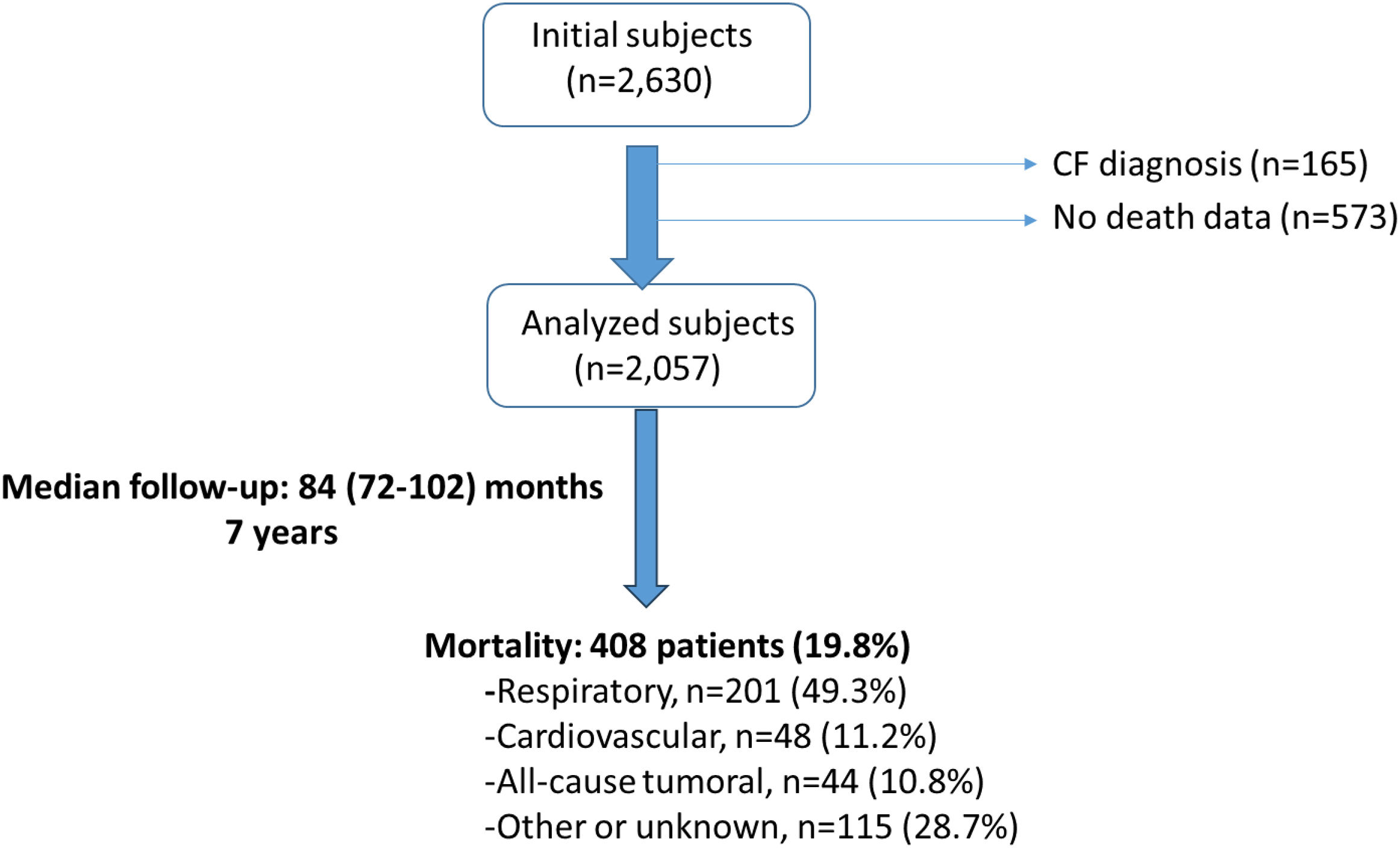
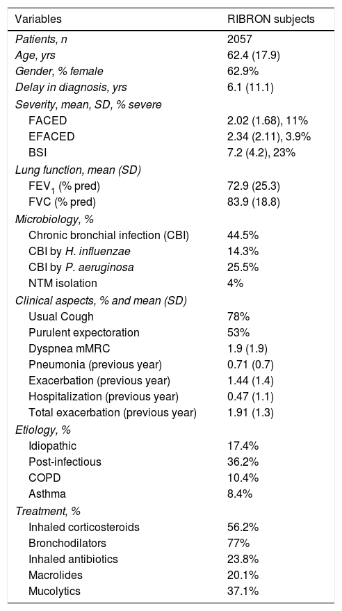
ResultsFrom the initial 2630 patients, 2057 were finally included in the study after excluding 165 patients with cystic fibrosis and 573 subjects without data on their vital status (Fig. 1). Mean age was 62.4 (62.9% women). FEV1 pred was 72.9% and 25.5% with CBI by PA. Baseline characteristics of included patients are shown in Table 1.

Baseline characteristics of included patients.
| Variables | RIBRON subjects |
|---|---|
| Patients, n | 2057 |
| Age, yrs | 62.4 (17.9) |
| Gender, % female | 62.9% |
| Delay in diagnosis, yrs | 6.1 (11.1) |
| Severity, mean, SD, % severe | |
| FACED | 2.02 (1.68), 11% |
| EFACED | 2.34 (2.11), 3.9% |
| BSI | 7.2 (4.2), 23% |
| Lung function, mean (SD) | |
| FEV1 (% pred) | 72.9 (25.3) |
| FVC (% pred) | 83.9 (18.8) |
| Microbiology, % | |
| Chronic bronchial infection (CBI) | 44.5% |
| CBI by H. influenzae | 14.3% |
| CBI by P. aeruginosa | 25.5% |
| NTM isolation | 4% |
| Clinical aspects, % and mean (SD) | |
| Usual Cough | 78% |
| Purulent expectoration | 53% |
| Dyspnea mMRC | 1.9 (1.9) |
| Pneumonia (previous year) | 0.71 (0.7) |
| Exacerbation (previous year) | 1.44 (1.4) |
| Hospitalization (previous year) | 0.47 (1.1) |
| Total exacerbation (previous year) | 1.91 (1.3) |
| Etiology, % | |
| Idiopathic | 17.4% |
| Post-infectious | 36.2% |
| COPD | 10.4% |
| Asthma | 8.4% |
| Treatment, % | |
| Inhaled corticosteroids | 56.2% |
| Bronchodilators | 77% |
| Inhaled antibiotics | 23.8% |
| Macrolides | 20.1% |
| Mucolytics | 37.1% |
NTM: non-tuberculous mycobacteria; mMRC: modified Medical Research Council; COPD: chronic obstructive pulmonary disease; BSI: Bronchiectasis Severity Index.
During the follow-up, the exacerbation annual rate was mean (SD): 1.86 (1.5), the hospitalization rate was 0.53 (1.2), and the total exacerbation rate was 2.39 (1.4).
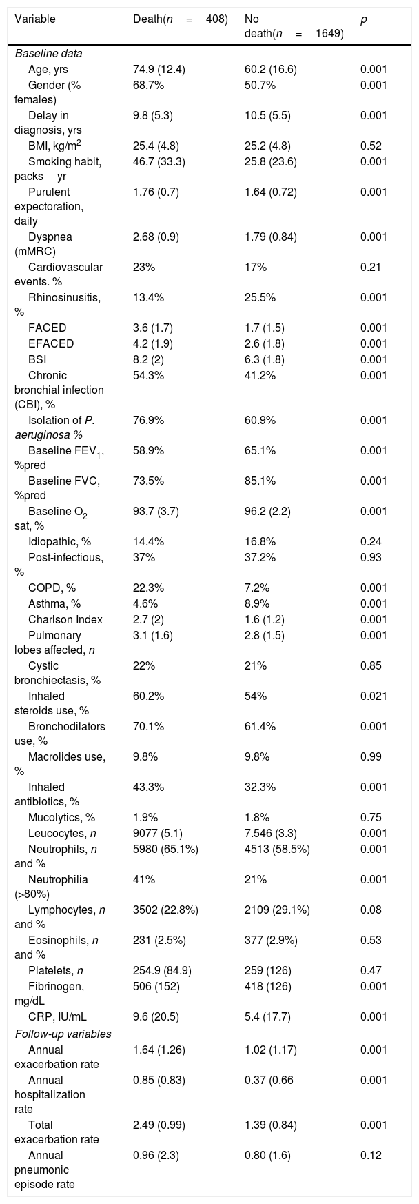
Univariable analysisTable 2 shows the comparison of baseline and follow-up variables between those patients alive and dead at the end of the study. There were 408 patient deaths during follow-up (19.8%). The deceased patients generally presented more severe bronchiectasis in almost all the domains that describe the disorder, as well as a greater number of comorbidities. Patients were followed up for a median (interquartile range) of 84 (72–102) months. The causes of death were respiratory (n=201; 49.3%), cardiovascular (n=48; 11.2%), malignant (n=44; 10.8%), and others or unknown (n=115, 28.7%).
Comparison between dead and alive subjects at the end of follow-up.
| Variable | Death(n=408) | No death(n=1649) | p |
|---|---|---|---|
| Baseline data | |||
| Age, yrs | 74.9 (12.4) | 60.2 (16.6) | 0.001 |
| Gender (% females) | 68.7% | 50.7% | 0.001 |
| Delay in diagnosis, yrs | 9.8 (5.3) | 10.5 (5.5) | 0.001 |
| BMI, kg/m2 | 25.4 (4.8) | 25.2 (4.8) | 0.52 |
| Smoking habit, packsyr | 46.7 (33.3) | 25.8 (23.6) | 0.001 |
| Purulent expectoration, daily | 1.76 (0.7) | 1.64 (0.72) | 0.001 |
| Dyspnea (mMRC) | 2.68 (0.9) | 1.79 (0.84) | 0.001 |
| Cardiovascular events. % | 23% | 17% | 0.21 |
| Rhinosinusitis, % | 13.4% | 25.5% | 0.001 |
| FACED | 3.6 (1.7) | 1.7 (1.5) | 0.001 |
| EFACED | 4.2 (1.9) | 2.6 (1.8) | 0.001 |
| BSI | 8.2 (2) | 6.3 (1.8) | 0.001 |
| Chronic bronchial infection (CBI), % | 54.3% | 41.2% | 0.001 |
| Isolation of P. aeruginosa % | 76.9% | 60.9% | 0.001 |
| Baseline FEV1, %pred | 58.9% | 65.1% | 0.001 |
| Baseline FVC, %pred | 73.5% | 85.1% | 0.001 |
| Baseline O2 sat, % | 93.7 (3.7) | 96.2 (2.2) | 0.001 |
| Idiopathic, % | 14.4% | 16.8% | 0.24 |
| Post-infectious, % | 37% | 37.2% | 0.93 |
| COPD, % | 22.3% | 7.2% | 0.001 |
| Asthma, % | 4.6% | 8.9% | 0.001 |
| Charlson Index | 2.7 (2) | 1.6 (1.2) | 0.001 |
| Pulmonary lobes affected, n | 3.1 (1.6) | 2.8 (1.5) | 0.001 |
| Cystic bronchiectasis, % | 22% | 21% | 0.85 |
| Inhaled steroids use, % | 60.2% | 54% | 0.021 |
| Bronchodilators use, % | 70.1% | 61.4% | 0.001 |
| Macrolides use, % | 9.8% | 9.8% | 0.99 |
| Inhaled antibiotics, % | 43.3% | 32.3% | 0.001 |
| Mucolytics, % | 1.9% | 1.8% | 0.75 |
| Leucocytes, n | 9077 (5.1) | 7.546 (3.3) | 0.001 |
| Neutrophils, n and % | 5980 (65.1%) | 4513 (58.5%) | 0.001 |
| Neutrophilia (>80%) | 41% | 21% | 0.001 |
| Lymphocytes, n and % | 3502 (22.8%) | 2109 (29.1%) | 0.08 |
| Eosinophils, n and % | 231 (2.5%) | 377 (2.9%) | 0.53 |
| Platelets, n | 254.9 (84.9) | 259 (126) | 0.47 |
| Fibrinogen, mg/dL | 506 (152) | 418 (126) | 0.001 |
| CRP, IU/mL | 9.6 (20.5) | 5.4 (17.7) | 0.001 |
| Follow-up variables | |||
| Annual exacerbation rate | 1.64 (1.26) | 1.02 (1.17) | 0.001 |
| Annual hospitalization rate | 0.85 (0.83) | 0.37 (0.66 | 0.001 |
| Total exacerbation rate | 2.49 (0.99) | 1.39 (0.84) | 0.001 |
| Annual pneumonic episode rate | 0.96 (2.3) | 0.80 (1.6) | 0.12 |
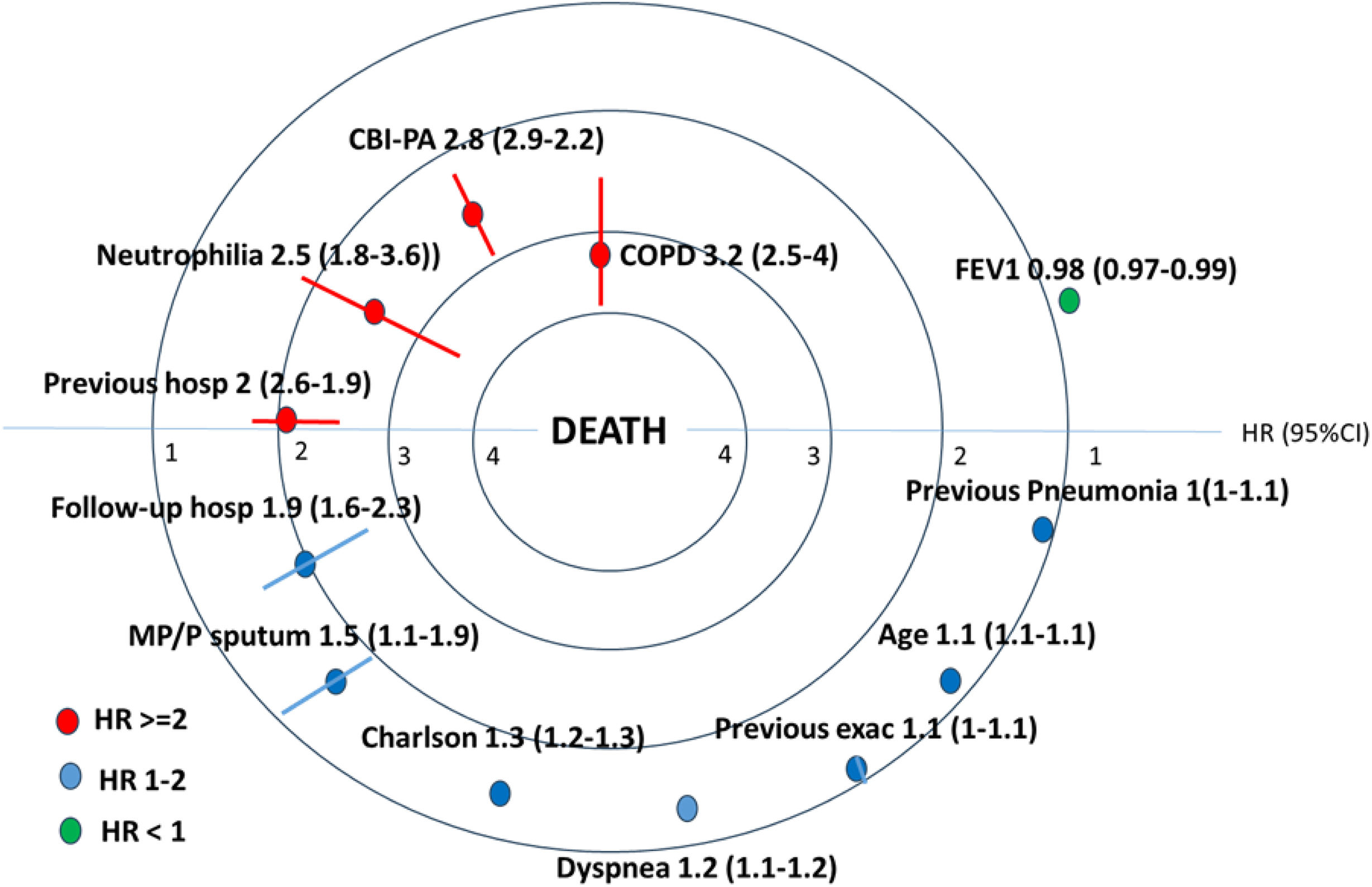
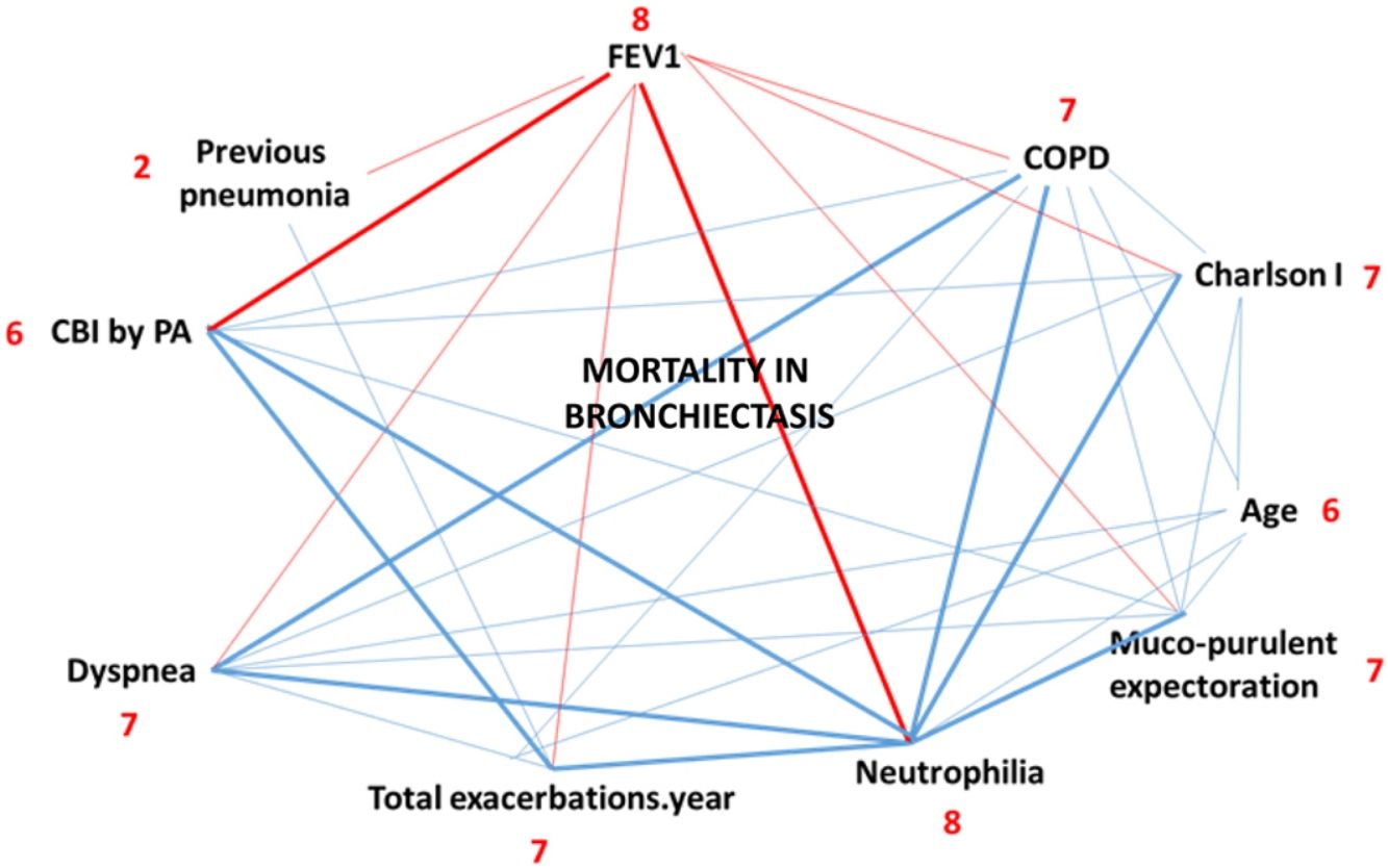
Thirteen variables were independently associated with mortality, as can be seen in Fig. 2, Table 1 Suppl. The strongest association with bronchiectasis mortality was the concomitant presence of COPD (HR 3.16; 95%CI: 2.5–3.9), the presence of CBI by PA (HR 2.57; 95%CI: 2.88–2.21), previous and follow-up hospitalizations (HR 2.01; 95%CI: 2.59–1.88 and HR 1.93; 95%CI: 1.63–2.32, respectively), as well as the presence of neutrophilia (HR 2.53; 95%CI: 1.81–3.61). Other variables such as baseline and follow-up exacerbations, other comorbidities, age, FEV1, dyspnea, and pneumonic episodes were also associated with higher bronchiectasis mortality. Only the presence of baseline peripheral neutrophilia appears as a new factor associated to mortality in bronchiectasis. When a separate analysis was performed for deaths of respiratory and cardiac origin, we observed that the three factors most strongly associated with respiratory mortality were the presence of COPD (HR 3.27; 95% CI: 2.53–3.91), chronic bronchial infection by P. aeruginosa (HR 2.42; 95% CI: 2.04–3.27), and neutrophilia>80% (HR 2.30; 95% CI: 1.21–2.41). In contrast, for cardiovascular deaths, the main associated factors were previous cardiovascular events (HR 4.17; 95% CI: 4.01–5.74), arterial hypertension (HR 3.15; 95% CI: 2.71–4.09), and diabetes mellitus (HR 2.17; 95% CI: 2.61–4.11). Further details of these analyses are presented in Supplementary Table 2.
Exacerbations
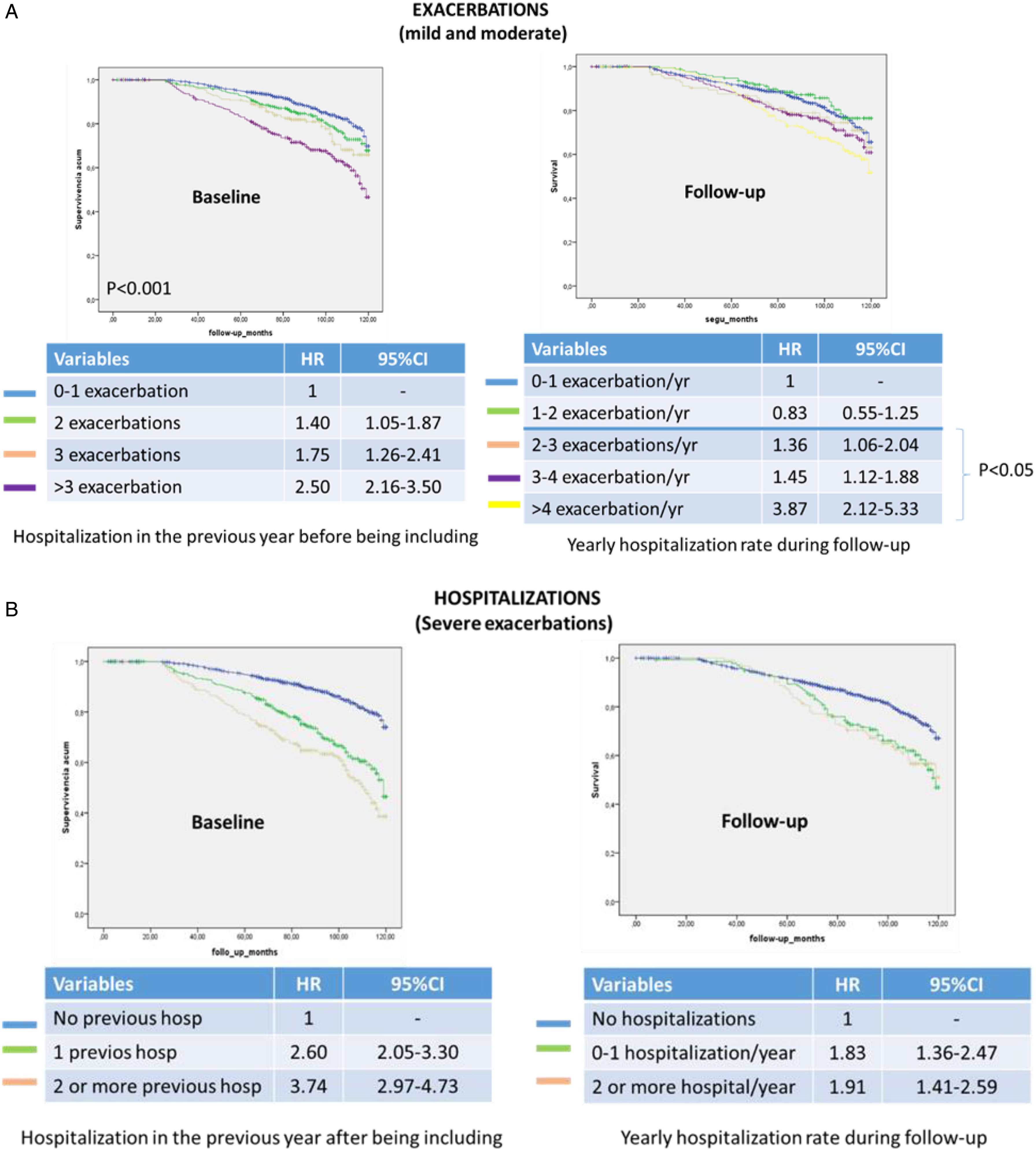
Fig. 3A shows a more detailed analysis of the exacerbations. There is a risk of greater mortality from mild or moderate exacerbations when there are two previous exacerbations or at least two exacerbations per year during the follow up, while, as regards hospitalizations (Fig. 3B), the presence of just one hospitalization previously or during the follow-up is sufficient to increase this risk by 2.60 and 1.83, respectively.
Impact of sputum characteristics, chronic bronchial infection by P. aeruginosa and coexistence of COPD
Both the presence of chronic mucopurulent or purulent sputum and a volume>20 cc per day were associated with higher mortality in bronchiectasis (Fig. 1 suppl).
On the other hand, the presence of CBI by PA was a strong factor in the relationship with mortality (HR 2.57; 96%CI: 2.88–2.21) as it has been in other studies (Fig. 2 suppl). However the coexistence of COPD was the strongest variable associated with long-term mortality in bronchiectasis, with an HR greater than three (3.16; 95%CI: 2.54–3.88) also seen in other previous studies (Fig. 3 suppl.)
DiscussionTo the best of our knowledge, this is the study with the largest number of included patients and the longest follow-up period published to date on mortality in bronchiectasis. According to our results, the coexistence of COPD, CBI by PA, baseline neutrophilia, and severe exacerbations (hospitalizations) at least doubles the probability of death in patients with bronchiectasis. The baseline multidimensional scoring system is a good predictor of long-term mortality, as well as being a means for dividing bronchiectasis into mild, moderate, and severe.
Although in the initial analysis LABA, inhaled antibiotics and use of macrolides in patients with bronchiectasis presented a significant risk of death, none of these variables were ultimately included in the final Cox model. The results of our study of factors related to increased mortality in bronchiectasis include those already published by Goeminne et al. [8] and Loebinger et al. [9], such as the presence of COPD, CBI due to PA, comorbidities, age, lung function, and symptoms such as the degree of dyspnea, as well as variables extracted from the studies of the BSI [11], FACED [12] and EFACED [13] scoring systems. Furthermore, in addition to achieving greater statistical power in the present study, due to the greater number of patients included and its longer duration, other variable such as the presence of neutrophilia (>80%), has emerged for the first time as risk factor.
The role of exacerbations deserves a section of its own. The severity and number of exacerbations have been shown to be factors related to death in bronchiectasis, especially when associated with CBI due to PA [2,3,11,13]. In our study, it was observed that in mild or moderate cases, the risk of increased mortality begins with two previous exacerbations per year or at least two exacerbations per year during the follow-up, while in the case of hospitalizations (severe exacerbations) the presence of just one hospitalization previously or during the follow-up is sufficient to increase this risk by 2.60 and 1.83, respectively.
Likewise, the presence of mucopurulent sputum and, more especially, chronic purulent sputum is associated with increased mortality. Furthermore, sputum color is the best biomarker of the severity of bronchiectasis, neutrophilic inflammation, and response to treatment [18].
For the first time, it has been observed that the presence of neutrophilia (percentage of neutrophils>80%) in peripheral blood is significantly associated with increased mortality in bronchiectasis (HR: 2.53 [95CI: 1.81–3.61]). This marker is very easy to obtain and probably reflects the increase in local inflammation of a neutrophilic nature, and therefore the biological activity of the disease in the already known vicious circle of inflammation and bronchial infection, especially through neutrophil elastases [19,20]. This opens an interesting window of prognostic and therapeutic opportunity with respect to the use of new anti-inflammatory drugs such as inhibitors of dipeptidyl peptidase 1 (DPP-1) (as has recently been demonstrated with Brensocatib) [21–26], and the potential role of other antibiotics with anti-inflammatory capacity, such as macrolides [27] and biologic treatments [28].
Among the strengths of the present study is that it is the study on bronchiectasis mortality with the greatest number of patients who died (n=408) and the longest duration (7 years), making it possible to establish robust relationships and find new factors that were hitherto unknown, based on a registry of 42 centers throughout Spain. Probably the main limitation of the study is that the vital status could not be determined in approximately 20% of the individuals; however, these patients did not show baseline characteristics different from those who were included in the study. Another limitation is that sputum volume was subjectively estimated by the patient using a graduated container in milliliters. Likewise, changes occurring during follow-up in terms of treatments or other variables were not taken into account, as only baseline variables were analyzed. Finally other limitation of the study is the need to take into account that these results must be validated in other countries with different characteristics of bronchiectasis patients and management settings, and that some of the diagnostic or therapeutic changes that occurred during the 7 years of follow-up have not been taken into account.
In conclusion, in our large series of patients with bronchiectasis, the variables most closely associated with mortality are the presence of COPD, CBI due to P. aeruginosa, severe exacerbations (hospitalizations), and the presence of peripheral neutrophilia.
Author's contributionsStudy design: MAMG, GO.
Data acquisition: RM, CO, RG, MG, LM, OS, LP, RG, JLRH, EB, CP, JRL, DR.
Data interpretation and writing the manuscript: MAMG, GO, MSDT.
Review of the first draft: RM, CO, RG, MG, LM, OS, LP, RG, JLRH, EB, CP, JRL, DR.
Review of the final draft: MAMG, GO, RM, CO, RG, MG, LM, OS, LP, RG, JLRH, EB, CP, JRL, DR.
All authors critically reviewed the manuscript, and approved its final, submitted, version.
Ethics approvalThe local ethics committee affiliated with the registry (number: 001-2012. Hospital Josep Trueta, Girona) approved the study and all the patients gave their written informed consent in their corresponding participating center.
Artificial intelligence useNo. The English translation was done by a native speaker (Matthew Clarke) with over 20 years of experience.
Data sharingData will be sharing after contact with the researchers and after approval by the authors of the present manuscript.
Funding statementInsmed Corporation (USA) granted this study without participation in any of the parts pf the research of this study.
Conflict of interestsAs we mentioned in the funding statement, Insmed Corporation (as we stated in the ScholarOne website) granted this study without participation in any of the parts of the research of this study, so authors declare no conflict of interest either with this work nor the corporation
Authors thank the participants in this study for their willingness to contribute to medical research.
The followings are the supplementary data to this article: