

Artificial intelligence (AI) is rapidly transforming respiratory medicine by enabling the integration and analysis of complex multimodal data, including imaging, physiological signals, electronic health records, and patient-generated information. This invited state-of-the-art review synthesizes current evidence on AI applications across chronic airway diseases, lung imaging, thoracic surgery and oncology, and sleep medicine and non-invasive ventilation, with a strong focus on clinically meaningful use cases. We highlight how AI can support earlier and more accurate diagnosis, refined phenotyping and endotyping, improved risk stratification, and more individualized therapeutic strategies. Representative applications include AI-assisted spirometry interpretation, multimodal exacerbation prediction, advanced quantitative imaging, perioperative risk modelling, wearable-based sleep apnoea detection, and AI-assisted respiratory support decision-making. Alongside these opportunities, we discuss cross-cutting challenges that currently limit routine implementation, including data quality and representativeness, insufficient external validation and generalizability, algorithmic bias, limited explainability of complex models, and concerns regarding privacy and data governance. Throughout the review, we argue that AI should be conceived as a tool to augment, rather than replace clinical judgment, embedded within human-in-the-loop frameworks that preserve transparency, fairness, and accountability. We conclude by outlining priorities for future research and policy, such as prospective clinical evaluation, multicenter validation in diverse populations, and closer collaboration between clinicians, data scientists, and regulators, which will be essential to translate the promise of AI into tangible improvements in respiratory outcomes and patient-centered care.
Artificial intelligence (AI) is rapidly reshaping respiratory medicine through the integration and analysis of increasingly complex multimodal data sources, rather than relying on isolated single-modality datasets. Advances in machine-learning (ML) and deep-learning (DL) models, together with the expansion of digital health infrastructures, have enabled a shift from largely descriptive and reactive care models toward more predictive, preventive, and personalized approaches [1–3]. Across a broad spectrum of respiratory diseases, including airway diseases [4], interstitial lung diseases [5], sleep disorders [6], and thoracic malignancies [7], AI is beginning to influence multiple stages of the clinical pathway, from early detection and phenotyping to therapeutic decision-making and outcome prediction [8]. Recent advances in foundation models and multimodal AI architectures are further accelerating this transition by enabling the integration of heterogeneous clinical information across diagnostic modalities.
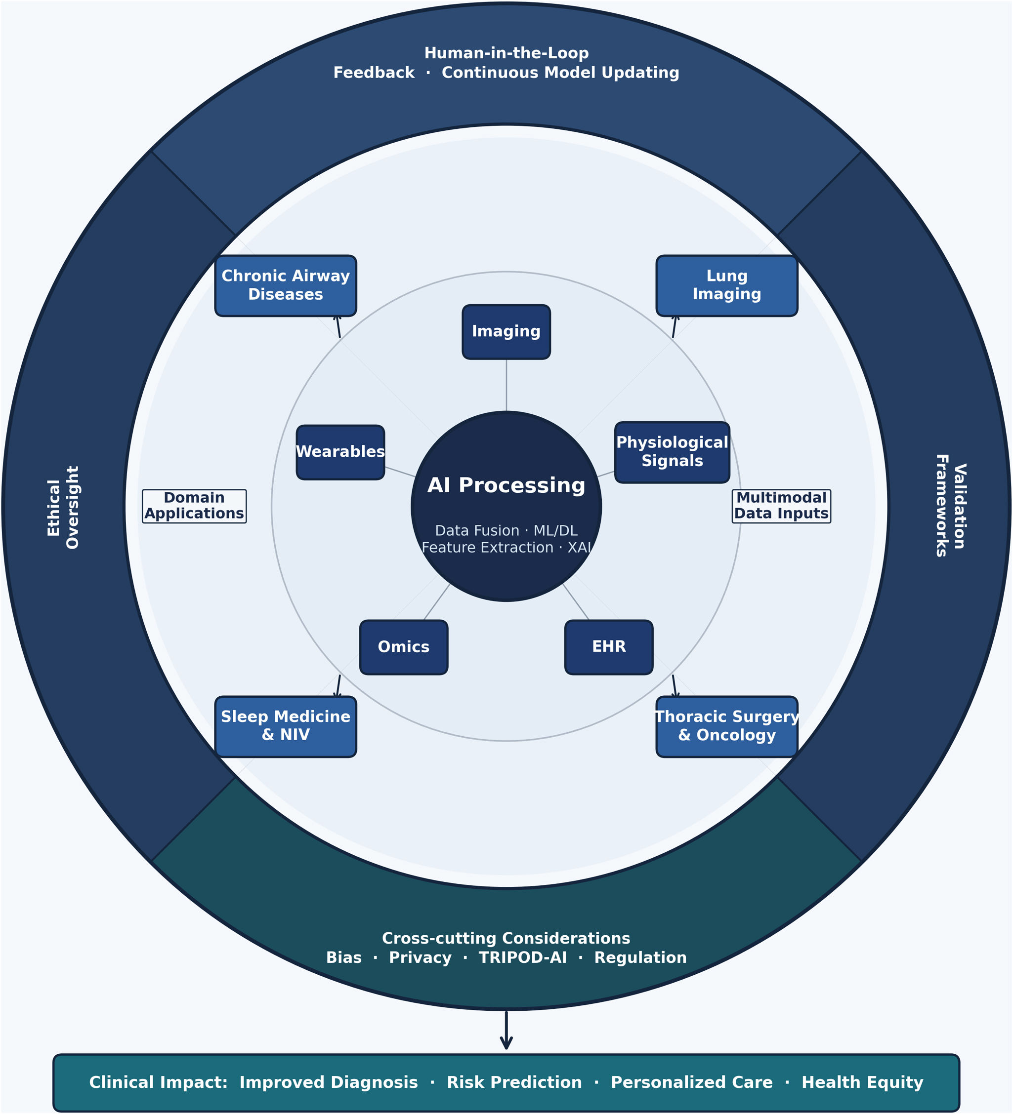
AI increasingly integrates diverse data sources (imaging, blood biomarkers, physiological signals, electronic health records [EHR], and diverse modalities of wearable and patient-generated data) to drive innovation [9]. This integration has the potential to improve diagnostic accuracy and resource utilization, but it also introduces important methodological, ethical, and, above all, implementation-related challenges [10,11]. This state-of-the-art review provides an integrated overview of the current applicability of AI across several domains of respiratory medicine, while also reflecting on the cross-cutting methodological and regulatory considerations that will determine whether these tools can be successfully translated into routine clinical practice [1]. Fig. 1 depicts the AI clinical pipeline as a concentric architecture in which five multimodal data input streams feed a central processing core that distributes domain-specific outputs across respiratory medicine, while governance considerations, including human-in-the-loop feedback, ethical oversight, and regulatory validation, continuously envelop all layers of the system.

Conceptual framework of artificial intelligence implementation in respiratory medicine. Artificial intelligence (AI) integrates multimodal data sources, including imaging, physiological signals, electronic health records (EHR), wearable devices, and omics, to support domain-specific applications across chronic airway diseases, lung imaging, thoracic surgery and oncology, and sleep medicine/non-invasive ventilation (NIV). Core AI processing encompasses machine learning (ML), deep learning (DL), feature extraction, data fusion, and explainable artificial intelligence (XAI). The outer framework highlights the cross-cutting requirements for successful clinical implementation, including ethical oversight, validation frameworks, continuous human-in-the-loop feedback, and model updating, together with methodological and regulatory considerations such as algorithmic bias, privacy, reporting standards, and governance. These elements ultimately determine the translation of AI into clinically meaningful outcomes, including improved diagnosis, risk prediction, personalized care, and health equity.
This invited state-of-the-art review was conceived to provide an expert-led synthesis of current applications, methodological challenges, and translational considerations of AI in respiratory medicine. Rather than aiming to perform a systematic review of the literature, the objective was to integrate contemporary evidence with the clinical and scientific perspectives of internationally recognized experts in key domains of respiratory medicine where AI is increasingly influencing research and clinical practice.
To support the narrative synthesis, targeted literature searches were performed in PubMed/MEDLINE by the authors responsible for each domain-specific section. No publication-date restrictions were applied. Searches combined general terms related to AI, ML, DL, and digital health with disease- and specialty-specific keywords relevant to the corresponding sections, including chronic airway diseases (CAD), thoracic imaging, thoracic surgery and oncology, sleep medicine, and non-invasive ventilation (Table S1, Supplementary Section). Additional references were identified through manual review of bibliographies from key publications and relevant regulatory and methodological documents.
The selected domains were chosen because they represent areas in which AI has achieved varying degrees of methodological maturity and clinical translation, thereby providing a broad overview of both opportunities and challenges across respiratory medicine. Priority was given to systematic reviews, randomized controlled trials, multicenter studies, landmark methodological papers, consensus documents, regulatory frameworks, and recent publications with direct clinical relevance. Particular attention was paid to issues of external validation, generalizability, explainability, implementation challenges, and reporting standards for AI-based research, including TRIPOD+AI and related methodological guidance [12].
Final reference selection and interpretation were performed by the contributing authors according to their respective areas of expertise; thus, each section was drafted by internationally recognized experts in the corresponding subspecialty. The coordinating authors harmonized style, structure, and critical appraisal across sections to ensure consistency throughout the review. This approach reflects the intended nature of an invited state-of-the-art review, in which expert appraisal and contextualization of rapidly evolving evidence complement traditional literature synthesis. Consequently, while every effort was made to provide a balanced and comprehensive overview of the field, as in other narrative reviews in emerging technological fields [13,14], the methodology does not aim for exhaustive literature retrieval and may be subject to selection bias. The purpose is instead to offer a clinically relevant and critically informed perspective on the current state of AI in respiratory medicine and the barriers that must be overcome for successful clinical translation.
Cross-cutting principles for artificial intelligence implementationWhether AI fulfils its promise in respiratory medicine will depend less on its theoretical potential than on the quality of its practical implementation. Successful ML applications require large, high-quality, well-governed and interoperable datasets integrating diverse sources of clinical information [1]. The integration of heterogeneous sources, including imaging, laboratory data, physiological signals, environmental variables, and, very importantly, unstructured EHR text, expands the analytical capabilities of AI while simultaneously increasing methodological complexity [13,15]. Consequently, the value of AI depends not only on algorithmic sophistication but also on the quality, representativeness, and integration of the data on which these models are developed. Successful clinical translation also requires robust external validation and adherence to established methodological reporting standards [1,16].
These principles are largely independent of the specific respiratory disease being studied and therefore underpin all AI applications discussed throughout this review. Regardless of the application, AI should augment rather than replace clinical expertise [17]. Human oversight remains essential to ensure that algorithmic outputs are interpreted within the appropriate clinical context and incorporated into patient-centered decision-making.
AI in chronic airway diseases (CAD)CAD, such as chronic obstructive pulmonary disease (COPD), asthma and bronchiectasis, are highly prevalent diseases that have a very significant health, social and economic burden. AI can help to address their diagnosis, biological heterogeneity, and clinical management. Below, there is a brief description of some examples, albeit the potential applications of AI in the diagnosis, management and follow up is much larger [18].
Underdiagnosis, misdiagnosis and late diagnosis, are key challenges in the management of CAD since underdiagnosis simply but very importantly implies no treatment, misdiagnosis can lead to inadequate treatment, and late diagnosis to less efficient therapy [18]. AI addresses all three scenarios effectively. For instance, AI can extract information from EHR notations (e.g., smoking, prematurity, repeated infections, exposure to environmental toxins, family history, symptom descriptions and/or treatment patterns) that may signal undiagnosed patients with CAD [18]. Likewise, AI-based quality control of spirometry facilitates diagnostic accuracy and reduces misdiagnosis [19].
Beyond diagnosis, AI addresses CAD's biological complexity through advanced phenotyping. CAD are complex and heterogeneous diseases and have several biological mechanisms underlying them (i.e., endotypes) that may overlap [20]. To investigate them, identify potential biomarkers and to guide therapy in clinical practice, AI can investigate multi-layer omics communities of patients with different underlying endotypes but similar clinical presentation (phenotypes) [21].
AI supports CAD clinical management through multiple approaches, as well. First, wearable sensor technologies allow transmission of patient-reported outcomes, physiological, and functional data directly to the health care system through automation, web-based data entry, or phone-based data entry [22]. The large volume of data generated can be integrated by AI which will be able then to propose a comprehensive summary of a patient's health to the practicing clinician. These can include information on adherence and appropriate use of inhaled medication [23–25] as well as early detection of exacerbations allowing prompt intervention [26]. Second, AI bridges patients and healthcare systems through intelligent communication tools [27]. For example, trained chatbots or avatars can address patients’ home-based health questions [28] and provide tailored recommendations. This AI-supported approach can bring health care closer to the patient and avoid unnecessary consultations while supporting effective self-management [27].
A recent perspective from the Global Initiative for Chronic Obstructive Lung Disease (GOLD) has highlighted several aspects where AI can be of help in patients with COPD [29]. These include: (1) addressing the underdiagnosis of the disease by proactive case finding extracting information suggestive of COPD from EHR and identifying or CT scans by quantifying structural lung abnormalities associated with COPD, such as emphysema or airway wall thickening in individuals included in lung cancer screening programs. AI can also help with the interpretation of spirometry and even extract novel, hidden, high-dimensional features from the flow-volume spirometry loops. Finally, there may be AI-based diagnostic alternatives to spirometry, including the analysis of respiratory sounds and voice features, forced oscillometry, single breath nitrogen test, e-nose signals and electrical impedance tomography; (2) contributing to COPD management by addressing patients heterogeneity using imaging and biologic biomarkers; (3) facilitate the implementation in clinical practice of disease guidelines, not only those of COPD but also those of related multimorbidity; (4) allow remote patient monitoring and facilitating the interaction between the patient and the health care system; and, (5) aid to improve our ability to establish the prognosis of the patient.
AI in lung imagingThe application of computer tools to study thoracic disease on imaging has a long history, dating back to efforts to quantify emphysema on computed tomography scans [30]. Yet key advances in the last 15 years such as deep learning algorithms, the creation of large natural imaging datasets to train these tools, graphical processing units which are able to run deep learning models and the wider availability of large medical imaging datasets [31,32] have accelerated the rate of growth of image analytics.
In chronic respiratory disease, AI is increasingly playing a role in improving quantitation of disease severity, disease progression and disease subtypes. In COPD, deep learning algorithms are increasingly being deployed to quantify mucus plugs [33] which have been associated with progressive COPD phenotypes and increased exacerbation risk [34]. Given that a wide range of organs are visible on a thoracic computed tomography (CT), including the heart, bones, muscles and fat, it is now also possible to quantify the multiorgan disease [35] that is commonly seen in patients with COPD [36].
In interstitial lung diseases, quantitative image analysis has focused on the detection of patients at risk of fibrotic disease progression by quantifying airway abnormalities [37], fibrosis extent [38,39], vascular changes or detection [40] of a UIP/IPF pattern of fibrosis [41,42]. Quantification of change in fibrosis extent on longitudinal CT imaging has also been used to estimate disease progression [38,43], which has gained particular relevance with the INBUILD [44] and FIBRONEER-ILD [45] trials showing therapeutic benefits in non-IPF progressive pulmonary fibrosis.
CT scans of patients with bronchiectasis can now be analyzed with software to measure bronchi wall diameters to quantify lack of airway tapering, and mucus plug burden [46]. Objectively measuring changes in airway dilatation and mucus plugging will have an important role in future therapeutic trials following the success of the ASPEN trial [47], heralding the advent of new therapies for this previously relatively neglected chronic lung disease [48].
Another area where AI holds great promise is the field of early diagnosis of chronic lung diseases. This is becoming increasingly a priority both clinically and for therapeutic trials [49–51], and is now possible through the analysis of large datasets of longitudinal imaging acquired for other aims, such as lung cancer screening programs [52]. These programs are enriched for participants at high-risk for developing chronic lung diseases, but where many participants may still remain in a pre-clinical or asymptomatic phase of disease. The large volume of thoracic CT data accessible to researchers has enabled the development of foundation models [53] which by learning the representation of a CT scan can classify disease patterns with high accuracy. Foundation models could therefore in the coming years improve detection of early disease phenotypes, prior to the advent of lung function disturbance.
A broad spectrum of challenges, however, precludes the speedy deployment of AI tools into clinical workstreams. As well documented in recent reviews [54,55], these can include challenges in developing reproducible and generalizable AI models that are minimally affected by variations in image acquisition. Scan variability may result from differences in scanner models between study centres, differing image reconstruction kernels or patient factors such as breath hold variation that might change longitudinal disease quantification. Similarly, algorithms that work for a specific use case in one environment might not generalize to populations with differing disease burdens, for example a nodule detection algorithm to assess lung cancer risk developed in a European context might produce very different results if applied to setting where tuberculosis is endemic. Ensuring broad clinical uptake will require AI models to integrate smoothly into radiology workflows and in theory perform a variety of clinically meaningful tasks before a healthcare system considers investment into the technology worthwhile. Finally, developers and users of AI systems need clarity on how regulatory bodies may consider AI models that evolve and learn rather than remaining locked or frozen prior to clinical deployment [56,57]. This question becomes ever more exigent given the exponential advances of modern AI systems.
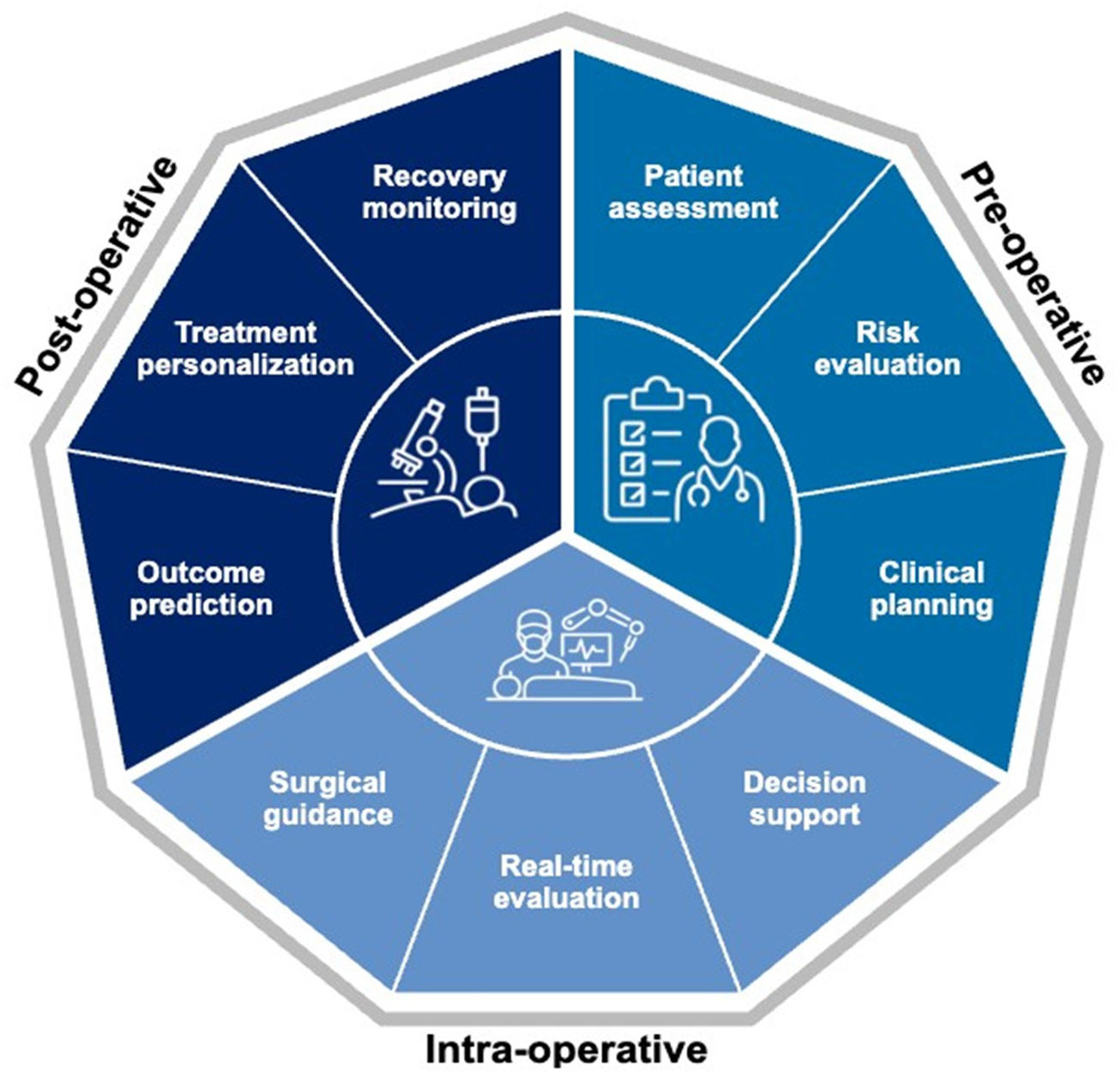
AI in thoracic surgery and oncologyAI is affecting thoracic surgical oncology practice by refining preoperative planning and increasing the precision of intraoperative guidance while carefully predicting postoperative outcomes (Fig. 2) [58–60]. Indeed, it has become customary to use ML, DL, computer vision, and robotic-assisted systems to optimize the clinical workflows of thoracic surgical patients [58,59]. As an example, the use of AI radiomics in the characterization of lung nodules provides higher AUC scores than LungRADS (in excess of 0.94) [61]. Thoracic surgeons are increasingly utilizing AI algorithms in the discrimination of malignant lung nodules as well as in the prediction of tumor stage (AUC 0.86–0.89) [62]. Moreover, the need for targeted therapies, either in a neoadjuvant or in an adjuvant setting, can be clarified by AI radiomics models that can detect the EGFR mutation status with an AUC ranging between 0.70 and 0.81 [63]. Conversely, the selection of candidates to upfront surgery can be refined by the prediction of recurrence risk [64] as well as an accurate prediction of postoperative complications after lung surgery with AUC values ranging from 0.73 to 0.90 [65–67]. The use of predictive AI models is justified by comparative studies with conventional risk scores showing a clear difference in performance expressed by AUC scores (0.734 vs 0.521 generated by the Charlson Comorbidity Index) [68]. In this setting, the personalization of perioperative management and the attendant patient counseling based on the above risk prediction models is becoming a reality [66,67].

Artificial intelligence applications across the thoracic surgery pathway. Schematic representation of current and emerging AI applications throughout the thoracic surgical continuum. Representative examples include radiomics-based pulmonary nodule characterization, prediction of histology and actionable genomic alterations, surgical risk assessment and planning, three-dimensional reconstruction, intraoperative navigation and robotic assistance, postoperative prediction of complications and recurrence, and longitudinal outcome monitoring. The figure summarizes the translational landscape described in the text and does not imply equivalent levels of evidence or implementation maturity across applications. Although several approaches have demonstrated promising diagnostic and predictive performance, most remain investigational and require prospective multicenter validation, demonstration of clinical benefit, and successful integration into routine clinical workflows before widespread adoption.
AI-driven technologies like 3D reconstruction, augmented reality navigation, and real-time intraoperative guidance systems are revolutionizing the surgical knowledge of anatomic structures and dissection planes, thereby facilitating the planning of challenging resections [58–60]. AI-enhanced robotic assisted thoracic surgery (RATS) can increase the precision of lung cancer surgery based on accurate hilar mapping which leads to the recognition of the least frequent anatomic variations [60,69]. AI-related technological advancements in robotic surgery will likely create further incentives towards training excellence while extending surgeons’ professional longevity [70,71]. The caveats that may hinder wider utilization and generalizability of AI algorithms include lack of sufficient study sample size, incompleteness of datasets, external validation, model calibration and mitigation of possible biases [64]. These potential drawbacks may affect the explainability of the logical process behind AI-models generated conclusions yielding the so-called Black Box problem [72]. In conclusion, despite the encouraging diagnostic and predictive performance reported across multiple applications, the current evidence warrants cautious interpretation. Many published AI studies in thoracic surgery and oncology remain retrospective in design, derived from single-center or highly selected datasets, and have undergone no or limited external validation. It remains uncertain whether improved model-performance metrics translate into meaningful clinical benefit, such as better patient outcomes, more efficient care delivery, or superior decision-making in real-world practice. Consequently, high discrimination performance alone should not be equated with clinical readiness, and further prospective, multicenter adequately powered validation studies are required before widespread implementation can be recommended [59,73,74].
AI and sleep medicine and non-invasive ventilationAmong all respiratory subspecialties, sleep medicine and home NIV may represent one of the earliest real-world ecosystems for AI implementation because both diagnostic and therapeutic devices are increasingly connected and continuously generate high-dimensional physiological data [75]. The continuous integration of wearable-derived physiological signals may also facilitate the development of digital biomarkers capable of detecting disease progression, treatment response, or impending respiratory decompensation.
In terms of diagnosis of obstructive sleep apnea (OSA), screening of EHR may be helpful to identify patients who are at high risk of OSA and related complications. Multimodal approaches could combine biomarkers, imaging, and screening tests to optimize diagnostic accuracy. Shin et al. [76] developed a multivariate model (with AUC=0.82) to predict OSA using BMI, pulmonary hypertension, hypertension and diabetes; however, at least in theory, a DL model using multimodal approaches could yield improved diagnostic accuracy and could perhaps detect disease before complications have already developed. Detection of sleep apnea using wearable AI was recently summarized in a meta-analysis by Abd-alrazaq [77]. This area is rapidly evolving with advances in wearable technology and analytical approaches. Retamales et al. developed an automatic system for sleep apnea estimation using publicly available data from home tests via abdominal belts, respiratory belts and/or pulse oximetry [78]. The authors were able to classify reasonably accurately 72.4% of patients into severities of apnea hypopnea index (AHI).
Counterfactual prediction models can estimate how outcomes in OSA might differ under alternative scenarios, such as changes in treatment adherence or the use of different therapeutic strategies. They are being developed in various areas of ML and specifically in respiratory science. These approaches allow consideration of “what if” scenarios in predicting the response to a future intervention [79,80]. Although the approach requires large data sets, the response to a therapy of a particular intervention can be anticipated a priori accounting for variability in treatment responses. Lu et al. recently reported a deep counterfactual inference model which allowed the comparison of response to high flow nasal cannula versus response to non-invasive ventilation for patients at risk of respiratory failure [79,80].
Unsupervised clustering of OSA may help to classify patients with varying disease characteristics. OSA is known to have variable endotypes (underlying mechanisms) as well as phenotypes (clinical expression of disease) [81,82]. Given that the one-size-fits-all approach to OSA has largely failed, the identification of groups who are likely to have differential responses to various interventions may help to guide future studies. As one example, Pepin et al. have reported health trajectories around noninvasive ventilation (NIV) initiation in clusters of patients with obesity hypoventilation syndrome (OHS). The authors defined marked heterogeneity in OHS and the use of NIV, the results of which had a major impact on clinical outcomes including up to tenfold differences in mortality between groups in various clusters [83].
OSA endotypes include anatomical compromise, upper airway dilator muscle function, ventilatory control instability (i.e. elevated loop gain), arousal threshold among others [81]. In theory agents to stabilize control of breathing such as oxygen or acetazolamide should benefit the subset of patients with OSA who have elevated loop gain as an underlying pathophysiological driver. However, loop gain per se is not a strong predictor of response to oxygen or acetazolamide therapy [84–86], leaving open the possibility that machine learning approaches could be used to identify the subset (or cluster) of OSA patients likely to respond to a particular intervention.
Beyond patient selection, AI may become increasingly relevant during long-term NIV follow-up. Modern ventilators continuously generate large volumes of telemonitoring data, including adherence, leak profiles, respiratory events, patient–ventilator interaction, and longitudinal physiological trends [8]. AI-based analysis of these data could support automated identification of patients at risk of treatment failure, detect evolving clinical deterioration before overt decompensation, facilitate individualized ventilator optimization, and prioritize patients requiring clinical review. Although these applications remain largely investigational, they illustrate how AI may transform home mechanical ventilation from reactive follow-up to proactive, data-driven care.
Overall, AI applications in sleep medicine and NIV are evolving rapidly, supported by increasingly connected diagnostic and therapeutic devices [75]. Nevertheless, most current applications remain at varying stages of methodological maturity, and robust prospective validation, demonstration of clinical benefit, and integration into routine workflows will be essential before widespread clinical adoption.
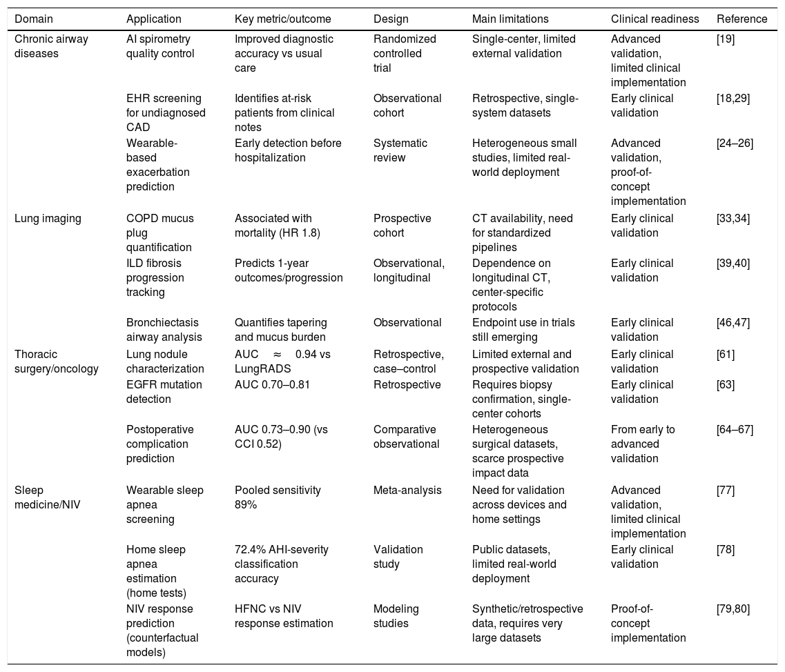
Table 1 integrates the representative AI applications discussed throughout the preceding sections, summarizing their reported performance, level of evidence, principal implementation barriers, and estimated stage of clinical readiness.
Key AI applications across respiratory medicine domains, including representative performance metrics, predominant evidence level, main implementation barriers, and estimated clinical readiness based on the predominant study design, extent of external validation, and degree of real-world implementation.
| Domain | Application | Key metric/outcome | Design | Main limitations | Clinical readiness | Reference |
|---|---|---|---|---|---|---|
| Chronic airway diseases | AI spirometry quality control | Improved diagnostic accuracy vs usual care | Randomized controlled trial | Single-center, limited external validation | Advanced validation, limited clinical implementation | [19] |
| EHR screening for undiagnosed CAD | Identifies at-risk patients from clinical notes | Observational cohort | Retrospective, single-system datasets | Early clinical validation | [18,29] | |
| Wearable-based exacerbation prediction | Early detection before hospitalization | Systematic review | Heterogeneous small studies, limited real-world deployment | Advanced validation, proof-of-concept implementation | [24–26] | |
| Lung imaging | COPD mucus plug quantification | Associated with mortality (HR 1.8) | Prospective cohort | CT availability, need for standardized pipelines | Early clinical validation | [33,34] |
| ILD fibrosis progression tracking | Predicts 1-year outcomes/progression | Observational, longitudinal | Dependence on longitudinal CT, center-specific protocols | Early clinical validation | [39,40] | |
| Bronchiectasis airway analysis | Quantifies tapering and mucus burden | Observational | Endpoint use in trials still emerging | Early clinical validation | [46,47] | |
| Thoracic surgery/oncology | Lung nodule characterization | AUC≈0.94 vs LungRADS | Retrospective, case–control | Limited external and prospective validation | Early clinical validation | [61] |
| EGFR mutation detection | AUC 0.70–0.81 | Retrospective | Requires biopsy confirmation, single-center cohorts | Early clinical validation | [63] | |
| Postoperative complication prediction | AUC 0.73–0.90 (vs CCI 0.52) | Comparative observational | Heterogeneous surgical datasets, scarce prospective impact data | From early to advanced validation | [64–67] | |
| Sleep medicine/NIV | Wearable sleep apnea screening | Pooled sensitivity 89% | Meta-analysis | Need for validation across devices and home settings | Advanced validation, limited clinical implementation | [77] |
| Home sleep apnea estimation (home tests) | 72.4% AHI-severity classification accuracy | Validation study | Public datasets, limited real-world deployment | Early clinical validation | [78] | |
| NIV response prediction (counterfactual models) | HFNC vs NIV response estimation | Modeling studies | Synthetic/retrospective data, requires very large datasets | Proof-of-concept implementation | [79,80] | |
EHR: electronic health record; CAD: chronic airway diseases; COPD: chronic obstructive pulmonary disease; CT: computed tomography; ILD: interstitial lung disease; EGFR: epidermal growth factor receptor; AUC: area under the curve; AHI: apnea–hypopnea index; NIV: non-invasive ventilation; HFNC: high-flow nasal cannula; CCI: Charlson Comorbidity Index.
The adoption of AI in respiratory care raises ethical, technical, regulatory, and societal challenges. For clarity, these divide into methodological limitations, arising from the models themselves and how they are built, and ethical, regulatory, and governance requirements, which determine whether a validated model can be deployed safely and equitably in routine practice.
Methodological limitationsOverfitting remains the central technical concern: An overfitted model learns dataset-specific noise rather than generalizable signal and may therefore fail when applied to new populations. Calibration problems are equally common. A model with high overall performance may still return misleading probabilities and often does so unevenly across clinical subgroups. Because such failures are silent at the point of care, they are a frequent make-or-break point: a model that discriminates well but is poorly calibrated for the local population will erode clinician trust the moment it is deployed.
Another related key concern is validation. Many published systems are still developed and tested in narrow, single-center conditions, leaving doubts about their generalizability [87]. Avoiding this requires rigorous external validation on multicenter datasets and adherence to reporting frameworks such as TRIPOD+AI and STROBE, which should be treated as minimum standards before any clinical claim [12]. Validation in real-life cohorts would be decisive: most tools are validated only retrospectively, whereas prospective assessment in unselected, routine-care populations is what demonstrates genuine clinical utility. A relevant example is provided by Kocks et al. [88]. An implementation that skips this step may perform well on paper yet perform poorly when introduced into routine clinical practice.
Explainability is a methodological requirement rather than an optional feature: for clinicians and patients to trust and challenge a prediction and possibly identify features that will not generalize well, its reasoning must be inspectable – and contestable. This is hardest for “black-box” architectures such as deep neural networks, but interpretability tools, for example, sensitivity-mapping methods such as NeuralSens, can make model behavior more transparent [89]. Where opacity cannot be reduced, adoption often stalls, since clinicians will not act on outputs they cannot interrogate. Methodological development should also move beyond purely associative prediction where appropriate, incorporating causal models that can inform intervention design, though these techniques remain relatively new and underexploited [90].
Ethical, regulatory, and governance requirementsUnder the EU AI Act, diagnostic and clinical-decision-support tools are classified as high-risk and subject to binding requirements on risk management, data quality, transparency, traceability, and human oversight; AI embedded in medical devices is already undergoing Notified Body conformity assessment and follows an extended timeline into 2027 [91].
Two further requisites operate. The first is post-market surveillance, the continuous performance monitoring mandated by the AI Act, essential to detect data drift, the progressive divergence between training data and the data a model later encounters, or concept drift, where the input–output relationship itself changes [92]. Without it, a model that passed validation can degrade unnoticed into a source of harm. The second is professional liability: when an AI system contributes to a clinical decision, responsibility must be clearly allocated, and the revised Product Liability Directive extends defective-product liability to software and AI, reinforcing rather than replacing the clinician's duty of oversight [93]. Unresolved liability is a well-recognized brake on adoption.
Algorithmic bias and equity are a foremost ethical concern: automatic does not mean objective, and even the choice of optimization target can embed bias – in asthma, for instance, children, parents, and clinicians may reasonably prioritize different outcomes, such as preserving long-term function versus avoiding attacks [94]. Such distortions are compounded by the underrepresentation of minorities in clinical research, exemplified by the exclusion of Black, smoking, and obese patients from studies of monoclonal antibodies for asthma [95]. Accordingly, the AI Act requires providers of high-risk healthcare systems to govern training, validation, and test datasets across their lifecycle – documenting sources and preparation, assessing quality and suitability, identifying gaps and biases, and ensuring datasets are relevant, sufficiently representative, and as complete as possible for the intended population and use. A tool that is accurate on average but inequitable across subgroups can be rejected in practice regardless of its headline performance.
Finally, privacy and data governance must be built into the way healthcare AI is deployed. Health data should be protected through measures such as pseudonymisation, limited and recorded access, confidentiality requirements, secure storage, and deletion when the data are no longer needed. These safeguards must be applied together with the GDPR principles of using data only for a specific purpose, collecting only what is necessary, protecting privacy from the outset, and ensuring that each person can access only the minimum data required for their role.
Complementing this, the European Health Data Space establishes a harmonized framework for the secondary use of health data, including algorithm training and validation, through national health-data access bodies and data permits [96]. Such harmonization supports uptake, as models that assume harmonized inputs frequently break against fragmented, non-standardized hospital data.
Where lawful data access pathways are absent, projects may suffer. However, privacy-preserving approaches such as high-fidelity synthetic data and federated learning, sharing only model updates rather than raw patient data, can close data gaps while safeguarding confidentiality [97].
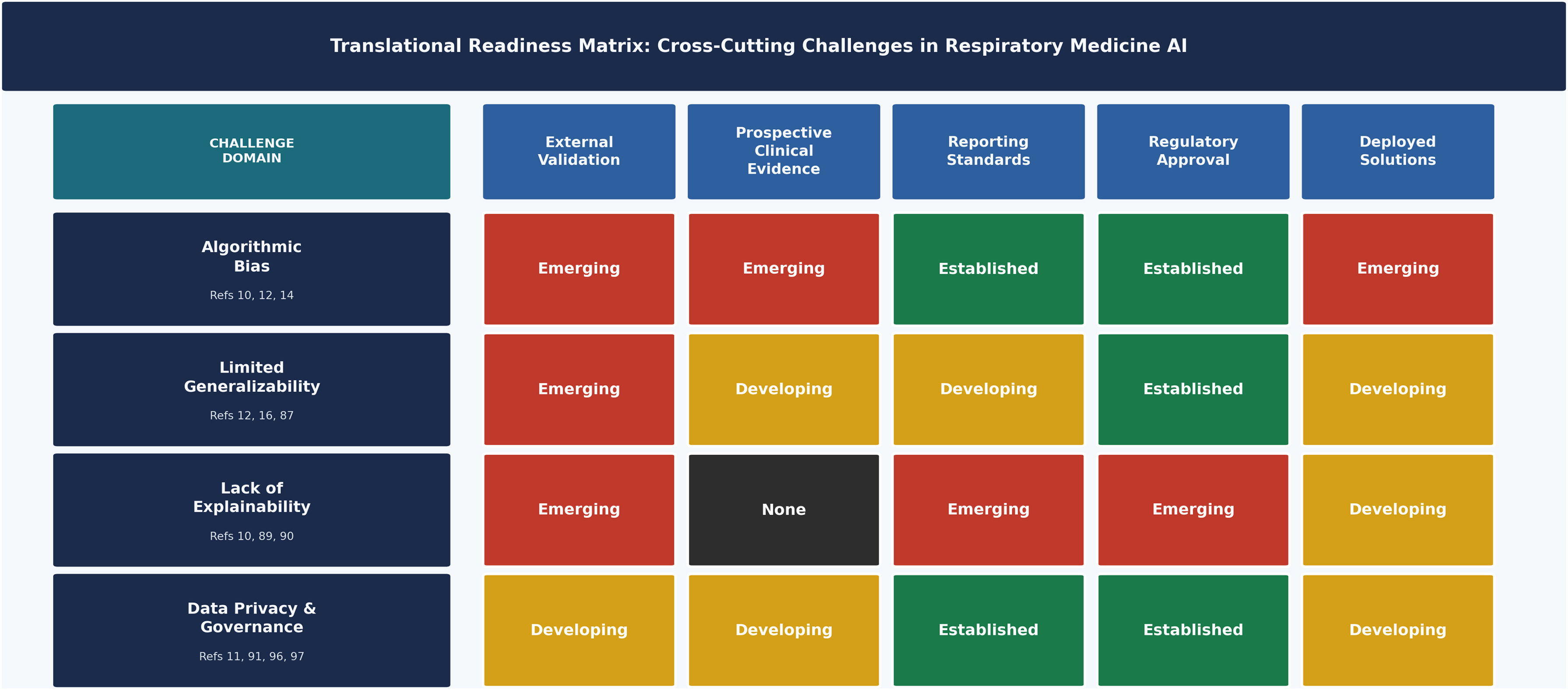
The cross-cutting challenges discussed above do not share the same degree of translational readiness. Fig. 3 maps their current maturity across five dimensions (external validation, prospective clinical evidence, reporting standards, regulatory approval, and deployed solutions) revealing a markedly uneven landscape. The barriers are not uniform, and the interventions required to overcome them differ in nature, urgency, and feasibility.

Translational readiness matrix of cross-cutting methodological and ethical challenges in respiratory medicine artificial intelligence. The matrix maps four challenge domains (rows) against five empirically grounded readiness dimensions (columns): external validation, prospective clinical evidence, reporting standards, regulatory approval, and deployed solutions. Each cell reflects a qualitative synthesis of the primary literature and is colour-coded according to a four-level maturity scale: None (black) – no published evidence identified in the respiratory AI literature; Emerging (red) – limited evidence from one or two pilot or single-centre studies; Developing (amber) – multiple studies available but with inconsistent results or incomplete implementation; Established (green) – systematic evidence base supported by operative regulatory frameworks or clinical guidelines. Supporting references are indicated at the row level. The maturity levels reflect the authors’ qualitative synthesis of the available literature reviewed in this article and are intended solely to illustrate the relative stage of clinical translation across domains rather than to provide quantitative comparisons or a formal evidence-grading system. The figure highlights the current imbalance between rapid technological innovation and the slower pace of prospective validation, regulatory approval, and routine clinical implementation, emphasizing that successful adoption of AI depends not only on algorithmic performance but also on robust evidence, transparent reporting, effective governance, and demonstration of real-world clinical benefit.
AI holds transformative potential for respiratory medicine by enhancing diagnostic precision, enabling sophisticated risk stratification, and facilitating truly individualized therapeutic strategies across diverse clinical domains. From chronic airway diseases, where AI improves spirometry interpretation and exacerbation prediction, to lung imaging applications that quantify disease progression and early phenotypes, thoracic surgery with refined preoperative planning, and sleep medicine through wearable-based apnea screening, these technologies are demonstrating tangible clinical utility, particularly in processing complex multimodal datasets that reveal patterns beyond human detection alone. However, translating this potential into routine practice will require several key steps, including the curation of high-quality, representative datasets, rigorous multicenter external validation, and effective strategies to reduce bias. Just as important are transparent model development, responsible data governance, and strong ethical oversight to support equitable and safe deployment without widening existing health disparities.
Future directions should emphasize prospective randomized controlled trials to validate AI-driven interventions (e.g., real-time NIV optimization or personalized COPD phenotyping), interdisciplinary collaborations between clinicians, data scientists, and ethicists, and standardized benchmarks for generalizability across global populations. AI must augment rather than supplant clinical judgment, maintaining human-in-the-loop oversight to balance innovation with patient-centered accountability. Only through methodological rigor, transparent governance, and sustained evaluation can AI translate its promise into measurable improvements in respiratory outcomes. The greatest challenge for AI in respiratory medicine is no longer developing increasingly sophisticated algorithms, but ensuring that these technologies become trustworthy, equitable, and clinically useful.
Authors’ contributionsDLP: Conceptualization, methodology, writing, original draft, figures, writing, review and editing. SL: Conceptualization, methodology, original draft, figures, writing, review and editing. DLP and SL contributed equally to this work. AA: Writing, review and editing (chronic airway diseases). JJ: Writing, review and editing (lung imaging). GR: Writing, review and editing (thoracic surgery and oncology). AM: Writing, review and editing (sleep medicine and non-invasive ventilation). JBS: Supervision, writing, review and editing. All authors reviewed and approved the final manuscript.
Declaration of generative AI and AI-assisted technologies in the writing processPerplexity Computer was used in the preparation of this manuscript exclusively for the conceptualization and graphical design of Figs. 1 and 3. No AI tool was used for writing, data analysis, or interpretation. All figures were reviewed, modified, and validated by the authors, who take full responsibility for the final content.
FundingThis work received no specific grant from any funding agency in the public, commercial, or not-for-profit sectors.
Conflicts of interestDaniel López-Padilla has participated in speaking activities, advisory committees, and consultancies from 2022 to 2026 sponsored by Air Liquide, AstraZeneca, Aerogen Ltd, Chiesi, FAES, Fisher&Paykel, Fundación Teófilo Hernando, GSK, Löwenstein, Nippon Gases, Philips, and Zambon; grant funding from Menarini and Aerogen Ltd. Sara Lumbreras and Àlvar Agustí have no conflicts of interest to disclose. Joseph Jacob declares consultancy fees from Boehringer Ingelheim, F. Hoffmann-La Roche, GlaxoSmithKline, Voiant, NHSX; advisory board fees from Boehringer Ingelheim, F. Hoffmann-La Roche; lecture fees from Boehringer Ingelheim, F. Hoffmann-La Roche, Takeda, Open Source Imaging Consortium; grant funding from GlaxoSmithKline, Wellcome Trust (209553/Z/17/Z and 227835/Z/23/Z), Microsoft Research, Gilead Sciences, Chan Zuckerberg Initiative (CZIF2024-009938). Gaetano Rocco declares royalties from Scanlan International and Intuitive; non-compensated advisory board. Atul Malhotra reports income from Eli Lilly, Livanova, Zoll, Sunrise, and Powell Mansfield; NIH funding; philanthropic donation from ResMed to UCSD; co-founder and equity holder in Clairyon (formerly Healciso). Joan Soriano has received pharmaceutical company grants from 2022 to 2026 from Chiesi, GSK, Linde, and Novartis via Hospital Universitario de La Princesa; speaking activities, advisory committees, and consultancies sponsored by Air Liquide, Almirall, AstraZeneca, Boehringer Ingelheim, CHEST, Chiesi, CNPT, ERS, FTH, Gebro, Grifols, GSK, IHME, Laminar Pharma, Linde, Lipopharma, Menarini, Mundipharma, Novartis, OMS/WHO, Pfizer, ResApp, RiRL, ROVI, SEPAR, SAPIO, Seqirus, WHO EUR, Takeda, and Zambon; JBS declares no funding from tobacco manufacturers or their affiliates.
The following are the supplementary data to this article: