


We report the case of a 67-year-old man, former smoker, referred for a flexible bronchoscopy (FB), in view of radiological findings indicative of lung cancer. A detailed study of the imaging tests showed the existence of a lung mass and increased density in both lateral walls of the middle third of the trachea and the main carina with no pathological uptake on PET, visible in the flexible bronchoscopy (Fig. 1). Given these findings and the appearance of the lesions on endoscopy, lung mass and tracheal samples were obtained to confirm the endoscopic suspicion of tracheobronchial amyloidosis vs neoplastic infiltration.
 Computed tomography and PET. (C and D) Raised erythematous hypervascular plaques in the right middle third of the trachea and main carina extending along the medial wall of the right main bronchus. (E and F) Presence of acidophilic deposits in the submucosa corresponding to amyloid substance after specific staining with Congo red and thioflavin T.")
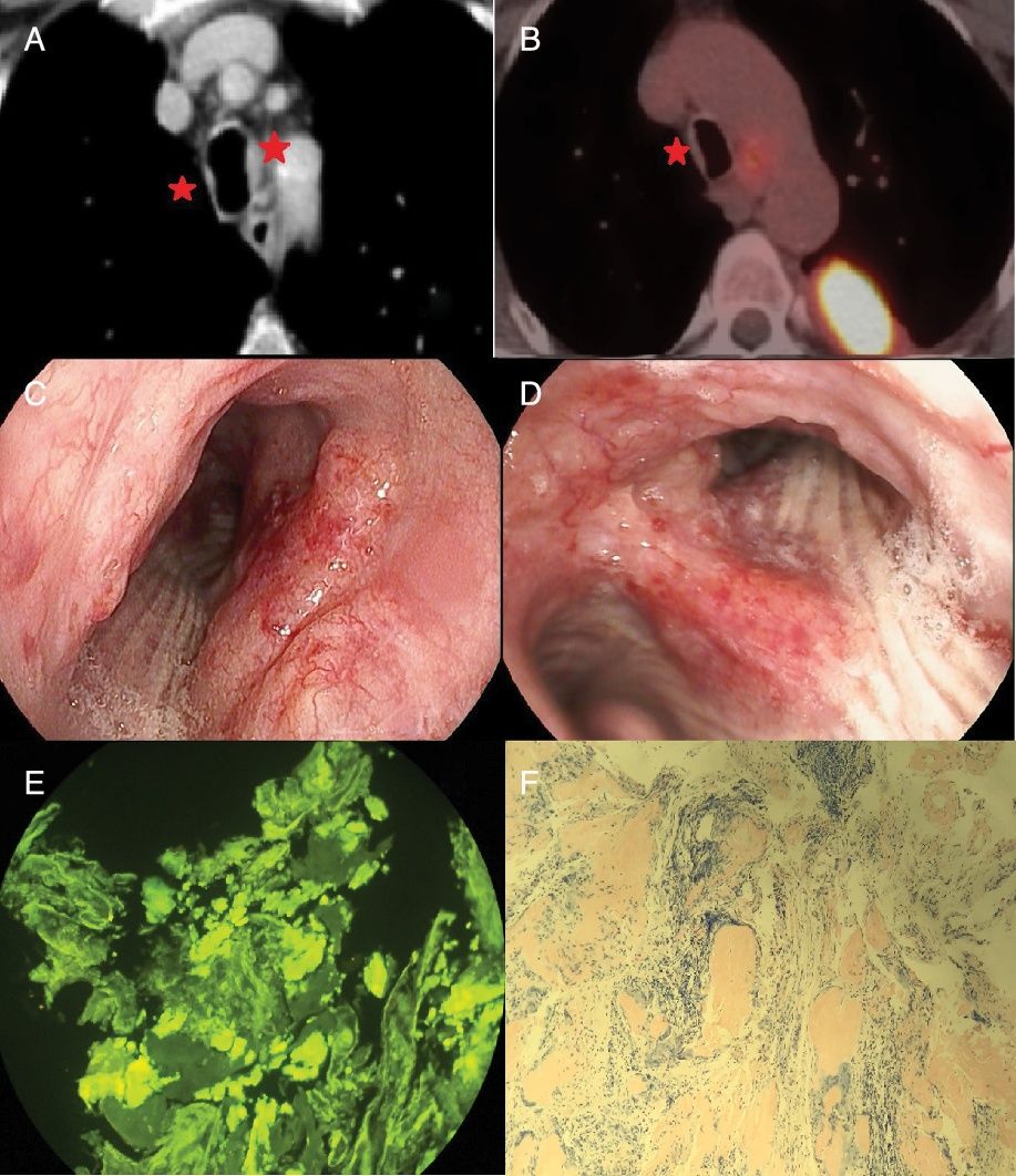
(A and B) Computed tomography and PET. (C and D) Raised erythematous hypervascular plaques in the right middle third of the trachea and main carina extending along the medial wall of the right main bronchus. (E and F) Presence of acidophilic deposits in the submucosa corresponding to amyloid substance after specific staining with Congo red and thioflavin T.
Histopathological studies established the diagnosis of localized primary tracheobronchial amyloidosis and surgically unresectable pulmonary adenocarcinoma (T2bN3M0). Tracheobronchial amyloidosis is a rare disease that, when detected on endoscopy, requires further studies to rule out secondary involvement.1 In the absence of a standardized treatment2 and the absence of tracheobronchial lumen compromise, chemotherapy was started. Endoscopic treatment was initially ruled out and the patient was followed up with endoscopic and clinical examinations. The presence of raised erythematous plaques in the tracheobronchial tree should prompt the endoscopist to consider this disease in the differential diagnosis.
Please cite this article as: de Vega Sánchez B, Disdier Vicente C, Martínez García G. Amiloidosis traqueobronquial como hallazgo incidental en un paciente con neoplasia pulmonar. Arch Bronconeumol. 2019;55:532.


