


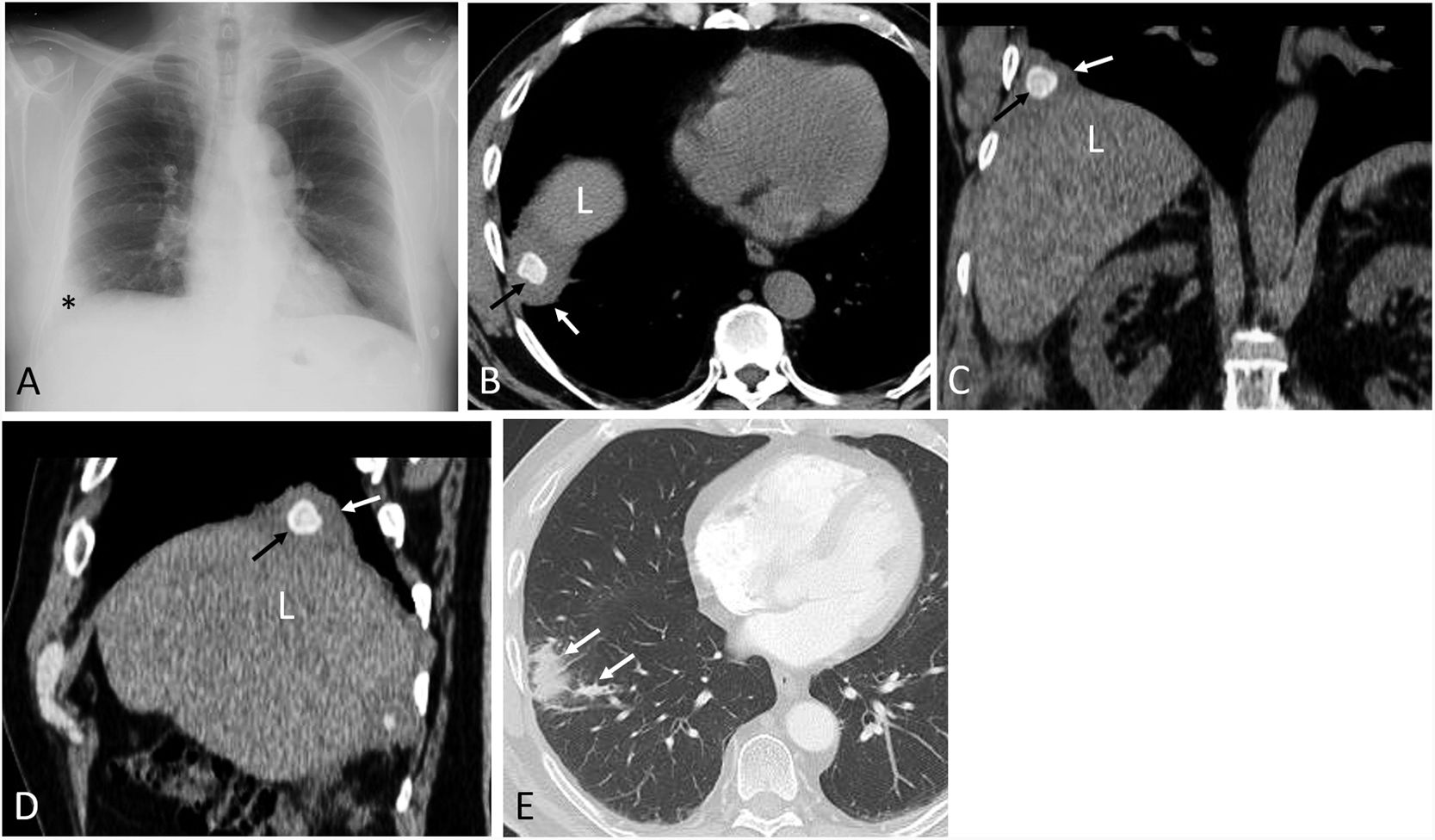
A 67-year-old patient presented to our hospital with cough and chest pain four months after undergoing a scheduled laparoscopic cholecystectomy for cholelithiasis. A chest radiograph showed a new-onset small right basal opacity and an ipsilateral costophrenic angle obliteration (Fig. 1A). Due to persistent chest pain despite analgesic treatment, a thoracic computed tomography (CT) was performed, which demonstrated a small right pleural effusion, a calcified polyhedral image within the ipsilateral pleural cavity, and inflammatory changes in the lateral basal segment of the right lower lobe (Fig. 1B–E). These findings were consistent with pleurolithiasis of biliary origin (PLBO) probably secondary to inadvertent intraperitoneal spillage of biliary content during laparoscopic surgery with posterior transdiaphragmatic migration. The patient underwent thoracoscopic removal of the pleural gallstone, lysis of adhesions, and partial resection of the lateral basal segment of the right lower lobe, with disappearance of his symptoms. No apparent diaphragmatic defect was demonstrated during surgery.
 Posteroanterior chest radiograph shows a small right basal opacity (asterisk). (B)–(D) Axial (B), coronal (C), and sagittal (D) thoracic CT images (mediastinal window) show a polygonal calcified lesion in the right pleural space (black arrow). Note the small pleural effusion (white arrow). “L” represents the liver. (E) Axial thoracic CT image (lung window) shows inflammatory changes in the right lower lobe (arrows).")
(A) Posteroanterior chest radiograph shows a small right basal opacity (asterisk). (B)–(D) Axial (B), coronal (C), and sagittal (D) thoracic CT images (mediastinal window) show a polygonal calcified lesion in the right pleural space (black arrow). Note the small pleural effusion (white arrow). “L” represents the liver. (E) Axial thoracic CT image (lung window) shows inflammatory changes in the right lower lobe (arrows).
Pleuroliths (or thoracoliths) are rare benign intrapleural calcified nodules that are usually incidentally found in the lower thoracic regions in imaging studies. The exact etiology of the majority of these calcified nodules is unknown, and mediastinal fat necrosis or pleural lipoma degeneration have been proposed as plausible mechanisms.1 PLBO secondary to gallstone spillage during surgery with posterior transdiaphragmatic migration is a very unusual complication and should be considered in patients with chest pain after laparoscopic cholecystectomy.2


