The diagnosis of multiple primary lung cancers (MPLC) accounts for approximately 0.2%–20% of all cases of lung cancer.1 The 2 presentations of MPLC, synchronous and metachronous, can manifest in 2 ways: either as a metastatic lesion of a primary lung tumor, or as multiple, unrelated lesions that can be defined as primary disease. The term MPLC is used to refer to the latter case, in which 2 or more primary malignant tumors are unrelated to each other, but present simultaneously or successively in the lung.2 Second lung lobectomies are very rarely described in the literature. The aim of this study was to determine postoperative morbidity and mortality in patients undergoing a second lung lobectomy. This is a descriptive study of postoperative morbidity and mortality (during hospitalization or in the first 30 days post-surgery) in a series of patients undergoing a second lung lobectomy between January 1, 2009 and December 31, 2014. Second lobectomies conducted to complete a pneumonectomy were not included in the analysis.
Of a series of 648 patients who underwent lung lobectomy, 6 (0.9%) received a second lobar resection. The study population consists of 4 men (66.6%) and 2 women (33.4%), mean age 53 years with a standard deviation (σ) of 7. Patient characteristics are listed in Table 1. The final approach was most often thoracotomy, although 50% of procedures began by video-assisted thoracoscopic surgery (VATS). Contralateral lobectomies were performed in 4 patients (66.6%). The major histological type, taking into account both surgeries, was pulmonary adenocarcinoma (75%) (9 results), the most frequent disease stage according to IASLC TNM classification (7th edition, 2009) in both surgeries was IA (75%); in 4 cases (66.6%), the disease was considered second primary lung carcinoma (SPLC), according to the criteria of Martini and Melamed.3 Morbidity after the second intervention was 50% (3 cases), with no postoperative mortality (Table 1).
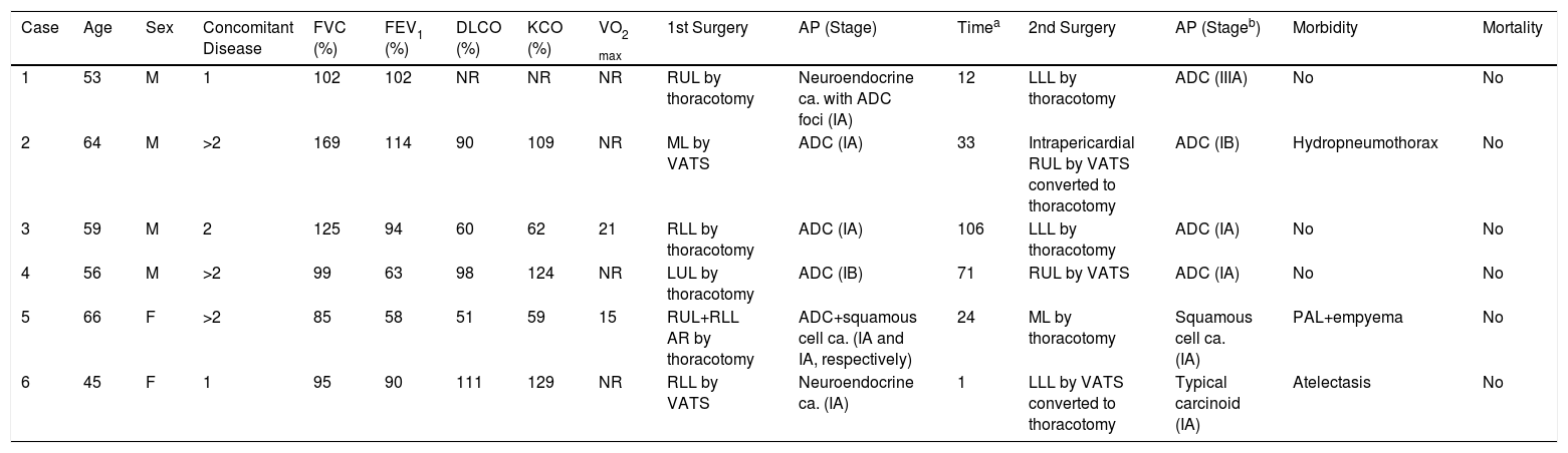
Age: At Second Surgery.
| Case | Age | Sex | Concomitant Disease | FVC (%) | FEV1 (%) | DLCO (%) | KCO (%) | VO2 max | 1st Surgery | AP (Stage) | Timea | 2nd Surgery | AP (Stageb) | Morbidity | Mortality |
|---|---|---|---|---|---|---|---|---|---|---|---|---|---|---|---|
| 1 | 53 | M | 1 | 102 | 102 | NR | NR | NR | RUL by thoracotomy | Neuroendocrine ca. with ADC foci (IA) | 12 | LLL by thoracotomy | ADC (IIIA) | No | No |
| 2 | 64 | M | >2 | 169 | 114 | 90 | 109 | NR | ML by VATS | ADC (IA) | 33 | Intrapericardial RUL by VATS converted to thoracotomy | ADC (IB) | Hydropneumothorax | No |
| 3 | 59 | M | 2 | 125 | 94 | 60 | 62 | 21 | RLL by thoracotomy | ADC (IA) | 106 | LLL by thoracotomy | ADC (IA) | No | No |
| 4 | 56 | M | >2 | 99 | 63 | 98 | 124 | NR | LUL by thoracotomy | ADC (IB) | 71 | RUL by VATS | ADC (IA) | No | No |
| 5 | 66 | F | >2 | 85 | 58 | 51 | 59 | 15 | RUL+RLL AR by thoracotomy | ADC+squamous cell ca. (IA and IA, respectively) | 24 | ML by thoracotomy | Squamous cell ca. (IA) | PAL+empyema | No |
| 6 | 45 | F | 1 | 95 | 90 | 111 | 129 | NR | RLL by VATS | Neuroendocrine ca. (IA) | 1 | LLL by VATS converted to thoracotomy | Typical carcinoid (IA) | Atelectasis | No |
ADC: pulmonary adenocarcinoma; AP: anatomical pathology; WR: wedge resection; ca.: carcinoma; DLCO: diffusing capacity of the lung for carbon monoxide; F: female; FEV1: forced expiratory volume in one second; FVC: forced vital capacity; KCO: diffusion constant for carbon monoxide; LLL: left lower lobectomy; LUL: left upper lobectomy; M: male; ML: middle lobectomy; NR: not recorded; PAL: persistent air leak; RLL: right lower lobectomy; RUL; right upper lobectomy; VATS: video-assisted thoracic surgery; VO2 max: maximal oxygen uptake.
The major intraoperative findings and technical difficulties in patients undergoing ipsilateral lobectomies were associated with firm pleural adhesions and hilar fibrosis, requiring the opening of the pericardium for adequate vascular control in 2 cases.
Median time between interventions was 24 months (range 1–106). After a mean follow-up of 203 weeks (3.5 years), 2 patients (33%) died due to disease progression, while the other patients are alive with no signs of relapse.
Survival outcomes after pulmonary resection for synchronous or metachronous cancers occurring less than 4 years apart are variable, but generally poor, suggesting that many of these patients may have had pulmonary metastases instead of SPLC. This situation justifies a thorough assessment of these patients to attempt to differentiate between metastasis and SPLC, although no distinguishing criteria have yet been defined.4 Most SPLC are of the same histological type and few criteria, including different histologic features or DNA breakpoints by sequencing, are reliable in themselves, so a thorough histological evaluation is required for a correct prognostic classification.5
The accurate assessment of these patients must also include stratification of the risk associated with pulmonary resection, focusing on the cardiologic evaluation, forced expiratory volume in one second (FEV1) diffusion capacity of the lungs for carbon monoxide, and cardiopulmonary exercise testing.6 In our setting, during the study period, the preoperative functional assessment protocol was based on the European Respiratory Society and European Society of Thoracic Surgeons 2009 guidelines on radical treatment of lung cancer, although 1 of our patients (case 1) did not meet the respiratory function criteria, due to limiting physical circumstances (patient with tracheostomy).
Due to the scant evidence, no specific recommendations are available on the role of adjuvant treatment in patients operated for operated for MPLC. In patients with synchronous MPLC, the tumor with the most advanced staging will determine the need for adjuvant treatment; the situation is unclear when a patient has 2 synchronous or metachronous stage I lesions, since, although the prognosis is worse than in patients with a single stage I NSCLC tumor, the absolute benefit of adjuvant cisplatin-based chemotherapy has not been demonstrated.7 Factors that could be considered when deciding whether or not to use adjuvant chemotherapy include the time interval between the cancers, certain unfavorable histologic characteristics (degree of differentiation, vascular and lymphatic invasion, solid and micropapillary pattern), tumor size, comorbidities, and the functional assessment of the patient.
Despite the limited number of patients in our series, we believe that a second lung lobectomy is a feasible technique, with zero mortality but with significant morbidity. Of the 3 cases that developed a complication, 2 patients had Grade III postoperative complications according to the Clavien-Dindo scale8 (complication that requires intervention without general anesthesia); and another had a Grade IV (life-threatening complication due to single organ dysfunction). To avoid major intraoperative bleeding in the event of an ipsilateral resection, complex dissection of the hilum should be anticipated, and intrapericardial dissection may be a surgical option to be taken into account in this group of patients.
Please cite this article as: Ordóñez Lozano PA, Royo Crespo I, Muñoz-González N, Embún Flor R. Segundas lobectomías pulmonares: ¿son factibles y seguras? Arch Bronconeumol. 2018;54:227–228.


