


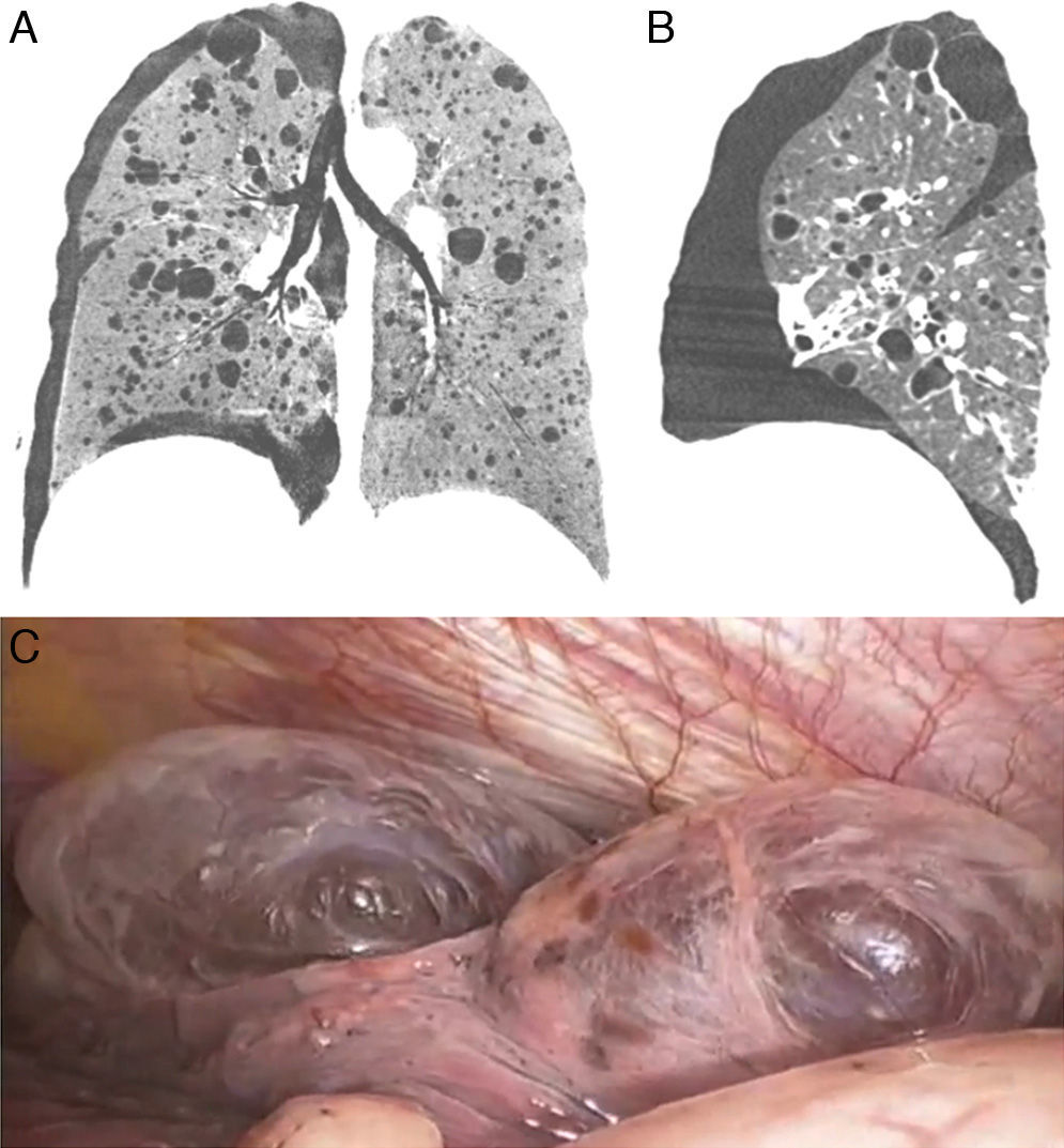
A 52-year-old woman presented with sudden onset of right chest pain and dyspnea. She had had two previous episodes of spontaneous pneumothorax. High-resolution computed tomography showed right pneumothorax and numerous thin-walled cysts in both lungs (Fig. 1a and b). Computed tomography also showed bilateral renal masses. Video-assisted thoracoscopic surgery (VATS) with bullectomy (Fig. 1c), and pleurodesis were performed. After 2 years, the patient remains asymptomatic.
 and sagittal (B) reformatted CT images show numerous bilateral, variably sized, thin-walled cysts (some subpleural) with right pneumothorax, consistent with lymphangioleiomyomatosis. During VATS, subpleural cysts in the lung parenchyma were noted (C).")
The lung cysts were compatible with lymphangioleiomyomatosis (LAM). The renal masses were diagnosed histologically as angiomyolipoma. Given these features, a diagnosis of tuberous sclerosis complex (TSC) was made.
TSC is an autosomal dominant disorder characterized by hamartoma formation in multiple organ systems. The definitive diagnosis can be made when the patient exhibits two major criteria (LAM, renal angiomyolipoma, cortical tuber, facial angiofibroma, retinal hamartomas, shagreen patch, subependymal nodule, ungual fibromas, among others) or 1 major and 2 minor diagnostic criteria.1,2
The clinical spectrum of TSC is wide, and many patients have minimal signs and symptoms. The occurrence of LAM in patients with TSC is associated with diffuse cystic lung lesions, and pneumothorax is a well-recognised complication. VATS may play an important role in the diagnosis and treatment of this condition.1,2
Please cite this article as: Pereira e Silva JL, Araujo Neto CA, Marchiori E. Neumotórax asociado a quistes pulmonares y masas renales. Arch Bronconeumol. 2016;52:106.


