


We report the case of a 48-year-old man, non-smoker, with a history of coronary disease, referred for the study of a lesion suggestive of pulmonary arteriovenous malformation (AVM) on chest computed tomography (CT), after a finding of right parahilar opacity on a chest radiograph.
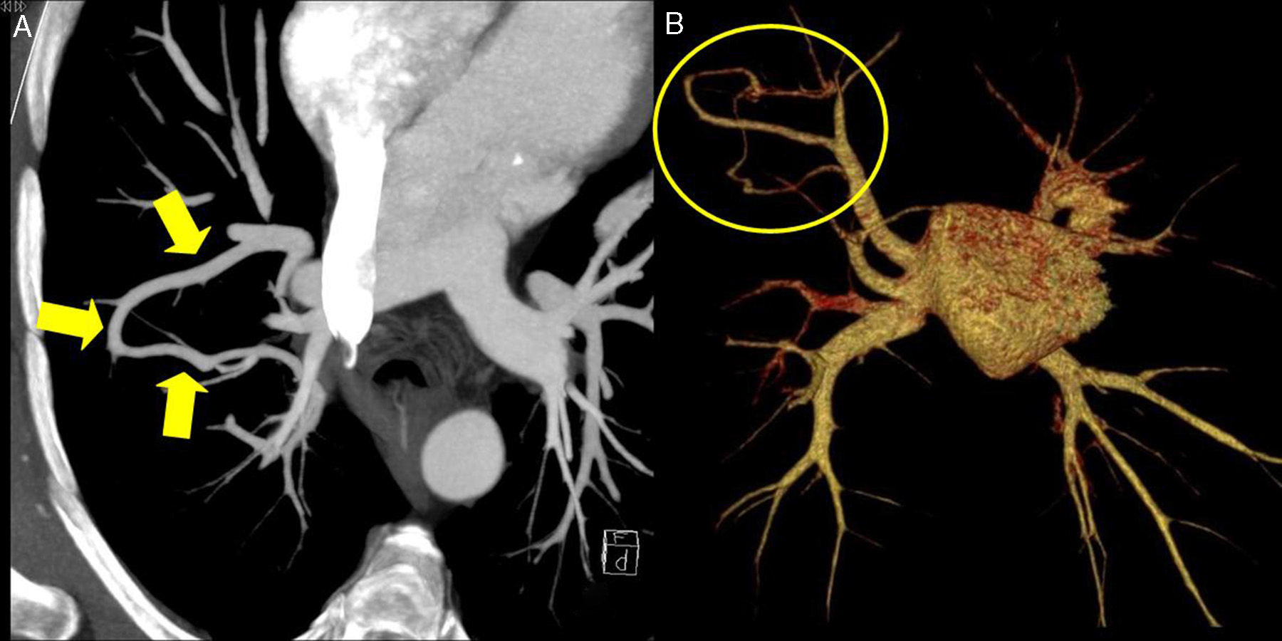
Transthoracic echocardiogram with contrast medium did not reveal right-to-left extracardiac shunt or any other abnormalities. Blood gases and spirometry were also normal. Pulmonary CT angiogram revealed pulmonary vein hypertrophy communicating the anterior segment vein with the posterior segment vein of the right upper lobe, suggestive of venous varix, draining to the right upper pulmonary vein.
Pulmonary venous varix is a rare entity, defined as a focal pathological dilation of a pulmonary vein, which may be isolated or associated with pulmonary venous hypertension, the latter mostly being due to mitral valve disease.1,2 It may mimic AVM on radiological tests, but a detailed examination of the images will reveal the connection with the venous vascular structures (Fig. 1), and the lack of arterial involvement. It is generally asymptomatic, as in our patient, and does not require treatment, but it should be differentiated from pulmonary arteriovenous malformation.
 Chest CT with maximum intensity projection (MIP) reconstruction, showing a tubular vascular structure communicating 2 pulmonary veins. (B) Chest CT with volume rendering (VR) reconstruction showing only the pulmonary veins and the left atrium, revealing the connection between the right pulmonary veins corresponding to the pulmonary venous varix.")
(A) Chest CT with maximum intensity projection (MIP) reconstruction, showing a tubular vascular structure communicating 2 pulmonary veins. (B) Chest CT with volume rendering (VR) reconstruction showing only the pulmonary veins and the left atrium, revealing the connection between the right pulmonary veins corresponding to the pulmonary venous varix.
Please cite this article as: Vaz N, Vollmer I, Perea RJ. Variz venosa pulmonar: una entidad rara que imita una malformación arteriovenosa. Arch Bronconeumol. 2016;52:562–563.


