


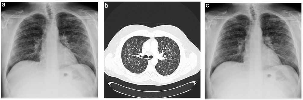
The patient was a 45-year-old man who was an active smoker and had no significant personal history. He consulted for constitutional symptoms, dyspnoea and two-month history of dry cough. A bilateral micronodular pattern respecting costophrenic angles was observed in the chest radiograph (Fig. 1A). Based on these findings, the study was expanded to include computed tomography (CT) of the chest. The CT scan revealed an interstitial pattern with heterogeneous microcystic changes in the upper lobes and also small centrilobular nodules that may cavitate, known as the Cheerio sign (Fig. 1B). Subsequently, a bronchoscopy was performed for bronchoalveolar lavage and transbronchial biopsy. The immunohistochemistry results showed CD45 and CD1 at less than 5%.
 Chest radiography showed a bilateral micronodular interstitial pattern. (B) CT showed a micronodular lung pattern with cavitation consistent with the Cheerio sign (blue arrowheads). (C) Chest radiography showed improvement at 6 months after smoking cessation.")
Despite the initial negative results, but high suspicion of histiocytosis, a surgical biopsy was requested, which revealed pulmonary Langerhans cell histiocytosis (PLCH).
PLCH is a rare interstitial disease that occurs in young adults and accounts for 3–5% of all adult diffuse lung diseases.1 This case is considered relevant because the diagnosis was made despite the negative immunochemistry results.
It is important to recognise this characteristic sign in the differential diagnosis of lung adenocarcinoma and metastasis or fungal, mycobacterial or rheumatoid nodules.2,3 This is because the therapeutic management of this condition is entirely different and early diagnosis can lead to a rapid recovery without medical treatment, as in our case with smoking cessation (Fig. 1C)
Authors’ ContributionsWe verify that all authors had access to the data and a role in writing the manuscript.
FundingThe authors declare that they have no funding.
Conflicts of InterestsThe authors state that they have no conflict of interests.


